Medical-diagnosis assisting apparatus, medical-diagnosis assisting method, and radiodiagnosis apparatus

A diagnostic aid and diagnostic device technology, applied in the direction of diagnosis, diagnostic recording/measurement, application, etc., can solve the problems of increased patient burden, prolonged examination time, and insufficient access

Inactive Publication Date: 2008-12-31
KK TOSHIBA +1
View PDF2 Cites 18 Cited by
  • Summary
  • Abstract
  • Description
  • Claims
  • Application Information

AI Technical Summary

Problems solved by technology

In addition, during examination / treatment, although contrast agent is injected and X-rays are irradiated to obtain 2-dimensional fluoroscopic images, sometimes sufficient information cannot be obtained depending on the angle, and it is necessary to change the irradiation direction and re-fluoroscopy every time, so the examination time is prolonged. , the burden on the patient (subject) has increased

Method used


Image

  • Medical-diagnosis assisting apparatus, medical-diagnosis assisting method, and radiodiagnosis apparatus
  • Medical-diagnosis assisting apparatus, medical-diagnosis assisting method, and radiodiagnosis apparatus
  • Medical-diagnosis assisting apparatus, medical-diagnosis assisting method, and radiodiagnosis apparatus

Examples


Embodiment 1

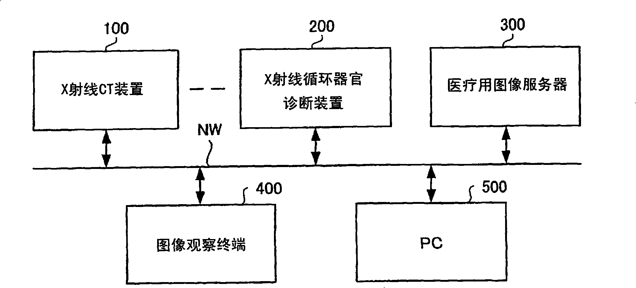
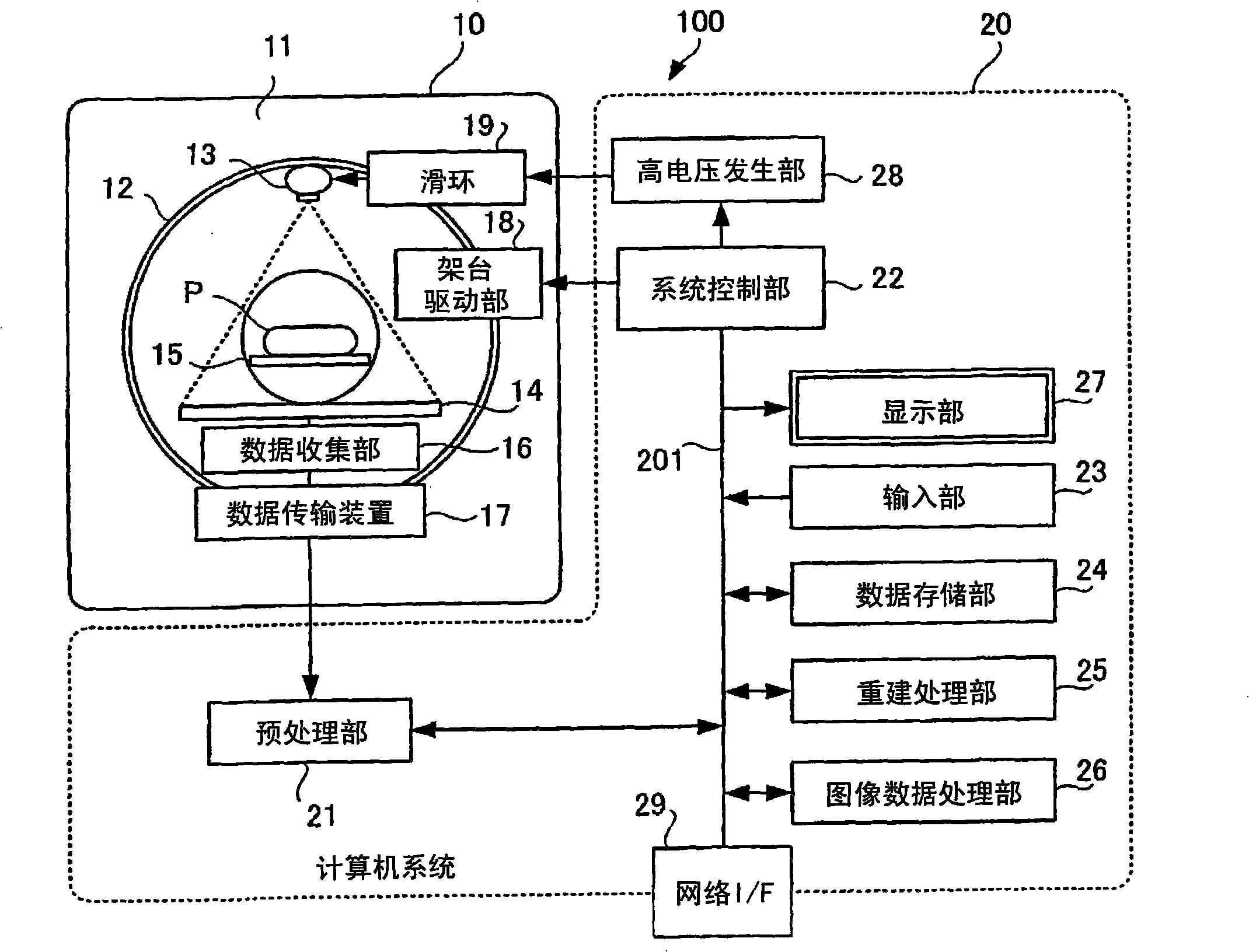
[0034] figure 1 A system configuration diagram showing a medical system using the medical diagnostic support device of the present invention. figure 1 The medical system has modalities such as an X-ray CT apparatus 100 and an X-ray circulatory organ diagnosis apparatus 200 as a radiation diagnostic apparatus. These modalities are connected to a network NW, and a medical image server 300 storing medical image information (including image data and incidental information) is connected to the network NW. Furthermore, an image viewing terminal (viewer) 400, an input / output terminal 500, and the like are connected to the network NW.

[0035] The X-ray CT apparatus 100 and the X-ray circulatory organ diagnosis apparatus 200 are used to image a subject to generate image data. Image data is stored in the medical image server 300 . In addition, the image observation terminal 400 is used to acquire and process image data and patient information stored in the medical image server 300 ,...

Embodiment 2

[0131] A second embodiment of the present invention relates to a radiation diagnostic apparatus having a guidance function in catheter inspection / treatment, and utilizes a part of the above-mentioned simulation function.

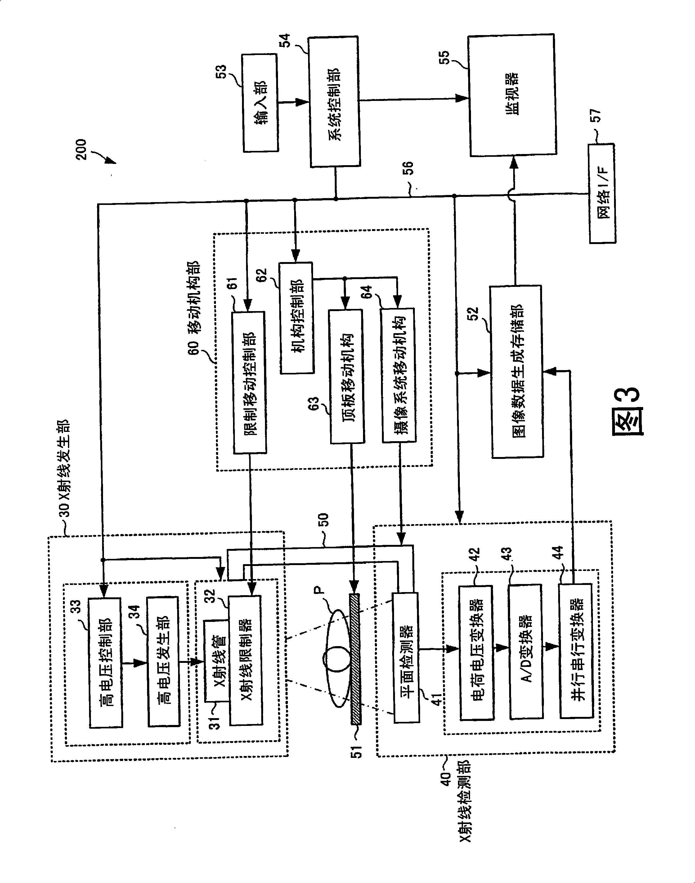
[0132] That is, a radiation diagnostic apparatus is provided which has a guidance function because the X-ray circulation organ diagnostic apparatus 200 includes the medical diagnosis support device 70 shown in FIG. 4 . In this case, the medical diagnostic assistance device 70 and storage device 81 shown in FIG. 4 are incorporated into the image data generation storage unit 52 of FIG. Device 55 is used concurrently.

[0133] In this embodiment, the actual fluoroscopic image obtained by the X-ray circulatory organ diagnostic apparatus 200 can be displayed, and the 3D image data collected and reconstructed by the X-ray CT apparatus 100 can be read in, and the CT value of the image data can be expressed as Arbitrary display angles display 3D images of coronary ...

Embodiment 3

[0147] In Embodiment 1, a simulation using a still image was described. However, the actual catheter operation is performed while watching a moving fluoroscopic image because the heart etc. is beating. Therefore, if a moving image is used instead of a still image in the simulation, a more realistic image can be provided.

[0148] In addition, during catheterization, a contrast agent is injected to facilitate visualization of blood vessels. Therefore, it is also possible to provide a more realistic image by simulating the change of the blood vessel image caused by the injection of the contrast medium. In addition, during the simulation, an MIP image assuming an X-ray fluoroscopic image when the catheter is operated is also displayed. Therefore, a more realistic image can be provided by simulating the change of the blood vessel image due to the injection of the contrast medium also on the MIP image.

[0149] Therefore, in Embodiment 3, a medical diagnosis support device that ...


PUM


Abstract

A medical-diagnosis assisting apparatus and a radiodiagnosis apparatus that include a simulation function prior to an examination / treatment, and a guide function during an examination / treatment are provided. Specifically, an extracting unit that extracts image data of a blood vessel portion to be an observation target from three-dimensional image data acquired by imaging a subject, a display unit that can display a three-dimensional image of the extracted blood vessel portion, a display-direction setting unit that displays the three-dimensional image of the extracted blood vessel portion on a display unit at a display angle specified by a user, and a simulation-image creating unit that simulates a course of a catheter when the catheter is to be inserted into the extracted blood vessel portion; and overlays a maker that indicates a position and a moving direction of the catheter on the three-dimensional image of the blood vessel portion are included.

Description

[0001] References to related applications [0002] This application enjoys the priority rights of Japanese Patent Application No. 2007-162673 filed on June 20, 2007 and Japanese Patent Application No. 2008-102782 filed on April 10, 2008. The entire content of this Japanese Patent Application will be published in cited in this application. Field of Invention [0003] The present invention relates to a medical diagnosis support device and a medical diagnosis support method. In medical diagnosis, especially when a catheter (catheter) is inserted into an object for examination and treatment of various diseases, a simulation display is displayed. ) image or guide (guide) image to assist the inspector, so that the safety is improved. In addition, it also relates to radiation diagnostic devices such as X-ray CT devices and X-ray circulatory organ diagnostic devices using medical diagnostic support devices. Background technique [0004] Conventionally, in medical systems using X-r...

Claims


Application Information

Patent Timeline
Patent Type & Authority Applications(China)
IPC IPC(8): A61B6/03A61B5/055
Inventor 小林秀明山形仁
Owner KK TOSHIBA
Why Eureka
Try Eureka