
Interfusion method of CT spacer and interest region capable of releasing CT image

A technology of region of interest and fusion method, which is applied in the field of fusion of CT tomographic images and CT positioning slices, can solve problems such as no solution, and achieve the effect of enriching information and expanding application value

Active Publication Date: 2009-05-06
NEUSOFT MEDICAL SYST CO LTD
View PDF0 Cites 12 Cited by
  • Summary
  • Abstract
  • Description
  • Claims
  • Application Information

AI Technical Summary

Benefits of technology

This technology allows physicians to see how well they are looking at certain parts inside their body by showing them with different types of x-ray films that help them identify specific areas within it. By combining these two techniques together, this can make better diagnoses than just analyzing one single type of data alone.

Problems solved by technology

Technological Problem: Current methods involve extracting specific areas of interests within 3D volume data that may help with diagnoses based solely upon visual inspection alone.

Method used


Image

  • Interfusion method of CT spacer and interest region capable of releasing CT image
  • Interfusion method of CT spacer and interest region capable of releasing CT image
  • Interfusion method of CT spacer and interest region capable of releasing CT image

Examples


Embodiment Construction

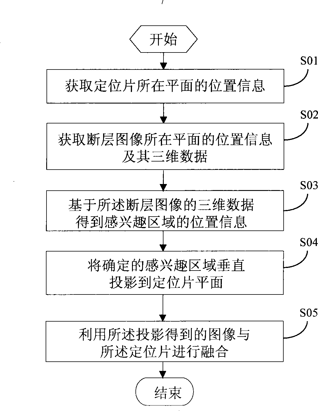
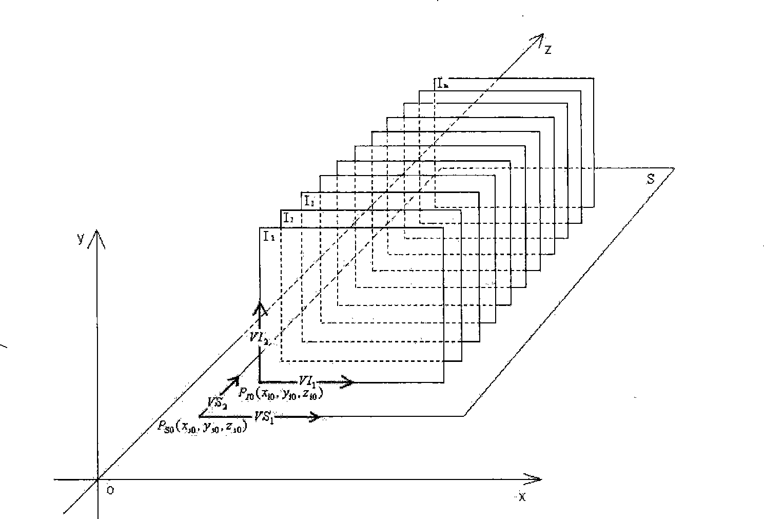
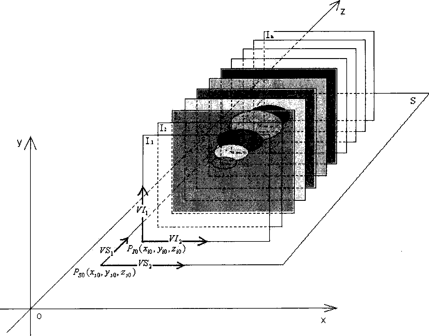
[0027] At present, for various diseases, examination methods based on CT equipment are becoming more and more popular. For the content of the present invention is to use the relationship between CT positioning film and CT tomographic scanning, the region of interest extracted in the tomographic sequence scanning image is fused and displayed on the CT positioning film image, and the image content of the CT positioning film is enriched, so that CT The positioning film can not only be used as a reference for the position of the tomographic scan, but at the same time, the doctor can observe the position information of the region of interest on the positioning film image in the anteroposterior or lateral projection, relative to other human tissues (such as bones), and provide more clinical diagnosis. much help.

[0028] In order to make the principles and characteristics of the present invention clearer, specific embodiments are accepted below for description.

[0029] refer to f...


PUM


Abstract

The invention discloses a method for realizing the fusion of an area-of-interest of a CT tomoscan and a CT locating card, which includes the steps: the position information of the plane in which the CT locating card is positioned is obtained; the position information and three-dimensional data of the plane in which the CT tomoscan is positioned are obtained; the position information of the area-of-interest is obtained based on the three-dimensional data of the tomoscan; the position information of the area-of-interest is utilized for projecting the area-of-interest on the plane of the CT locating card; and an image obtained from the projection is utilized and fused with the CT locating card. The extracted area-of-interest can be fused on the image of the CT locating card so as to provide richer information for the diagnosis of a doctor. To a certain extent, the method replaces the purposes of X ray films and expands the application value of the CT locating card.

Description


Claims


Application Information

Patent Timeline
Owner NEUSOFT MEDICAL SYST CO LTD
Learn More