Endoscopy device with removable tip

a technology of endoscope and tip, which is applied in the field of endoscope with removable tip, medical imaging, etc., can solve the problems of increasing the frequency of use of endoscope and increasing the risk of disease transmission

Inactive Publication Date: 2006-09-28
PERCEPTRONIX MEDICAL
View PDF13 Cites 159 Cited by
  • Summary
  • Abstract
  • Description
  • Claims
  • Application Information

AI Technical Summary

Problems solved by technology

The improvements in the design of endoscopes have caused an increase in the frequency of use of endoscopes, increasing the risk of transmission of disease as instruments are used and re-used.

Method used


Image

  • Endoscopy device with removable tip
  • Endoscopy device with removable tip
  • Endoscopy device with removable tip

Examples


Embodiment Construction

[0030] While preferred embodiments of the present invention are shown and described, it is envisioned that those skilled in the art may devise modifications of the present invention without departing from the spirit and scope of the invention.

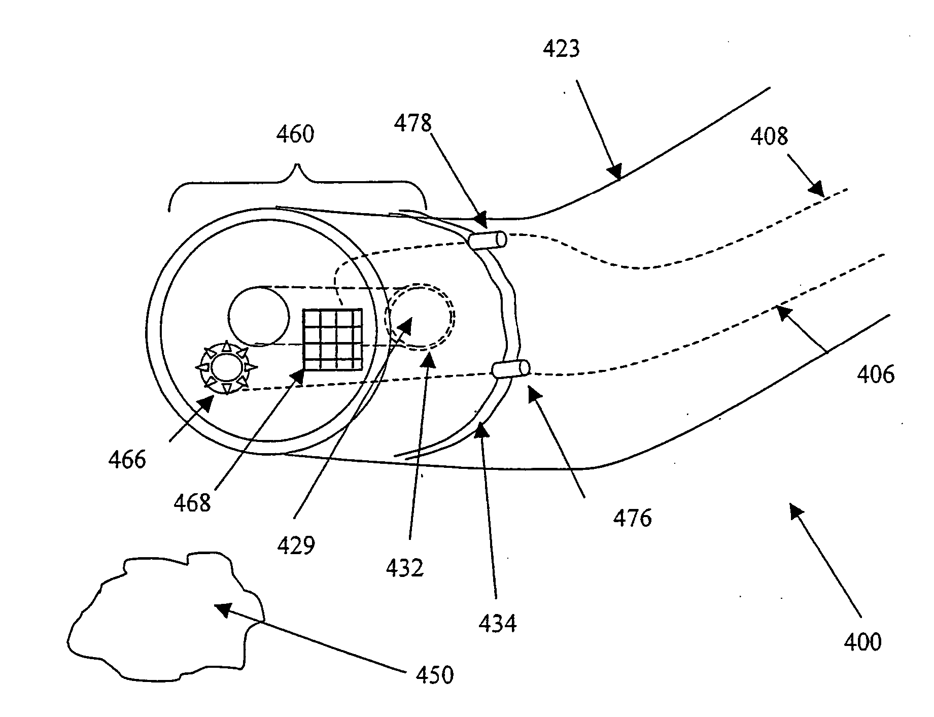
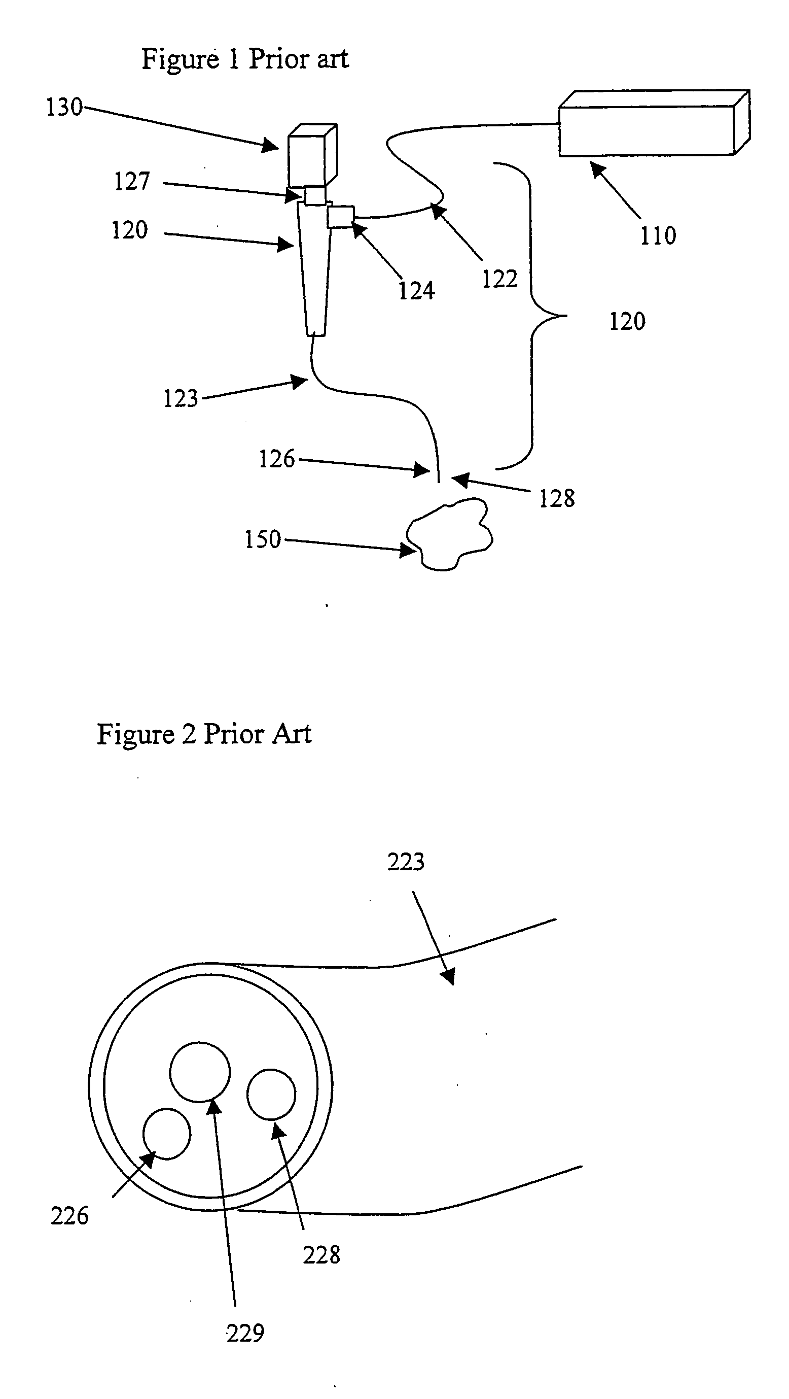
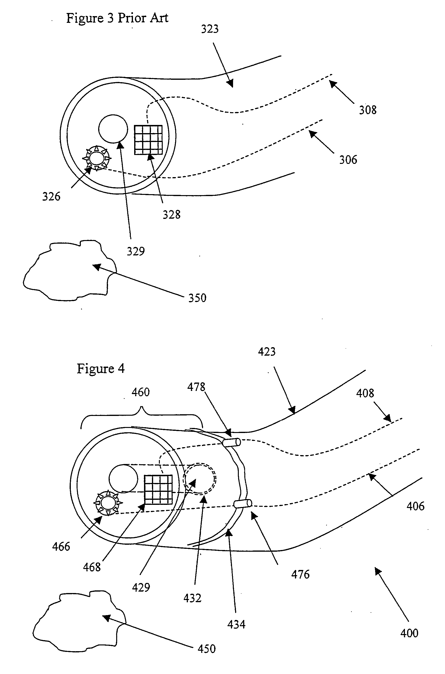
[0031]FIG. 1A shows an endoscope apparatus as is known in the prior art. Light source 110 generates interrogating radiation which can be some combination of white light for color images, narrow-band excitation light for fluorescence, or other narrow bands for spectral analysis or image normalization, or other types of light. Illumination source 110 provides light into endoscope assembly 120 via illumination fiber bundle 122 at junction 124. Illumination exits the distal end 123 of the endoscope 120, which in this example contains two fiber bundles 126, 128. Fiber bundle 126 directs illumination / excitation light to target tissue 150. The interrogating radiation is incident on a target 150, which will reflect, scatter, or fluoresce the interroga...


PUM


Abstract

The present invention provides an endoscope for in vivo imaging the cells, tissue, organs or body cavities of humans or other animals to observe and locate, diagnosis and / or treat disease. Illumination sources, image detectors, sensors may be provided alone or in combination on the removable tip allowing functional alterations or optimization for a particular procedure. Endoscope features such as an instrument channel supporting tissue sampling, suction, treatment, micro-surgery, optical computed tomography, confocal microscopy, laser or drug treatments, injections, gene-therapy, marking, implanting or other medical techniques are maintained.

Description

BACKGROUND OF THE INVENTION [0001] This invention relates to the field of imaging, especially medical imaging where an animal's cells, tissue, body cavities or organelles are accessed to facilitate surgery, treatment or to diagnose disease. [0002] Imaging is capturing electromagnetic radiation (wavelength and intensity) either reflected or emitted from an object of interest, in a manner which preserves or otherwise represents the spatial distribution of said radiation at the object. In the field of medical imaging, and more particularly endoscopy, light is utilized to illuminate body tissues and return a diagnostic or otherwise useful image. Historically, clinicians viewed white light reflectance (color) images through an ocular attached to the endoscope. More recently, with cost reductions and other computer advances, rather than viewing a tissue image through an ocular, endoscopic images are captured by an imaging device such as a video or digital camera and may be further process...

Claims


Application Information

Patent Timeline
Patent Type & Authority Applications(United States)
IPC IPC(8): A61B1/06A61B1/00
CPCA61B1/00105A61B1/053A61B1/00032A61B1/00101A61B1/0676
Inventor FERGUSON, GARY W.
Owner PERCEPTRONIX MEDICAL
Why Eureka
Try Eureka