
Inspection of tubular-shaped structures

A tube shape and structure analysis technology, applied in image data processing, instruments, calculations, etc., can solve problems such as time-consuming and error-prone, and achieve the effects of reduced user interaction, increased quantity, and reproducible measurement

Inactive Publication Date: 2010-03-24
KONINK PHILIPS ELECTRONICS NV
View PDF1 Cites 13 Cited by
  • Summary
  • Abstract
  • Description
  • Claims
  • Application Information

AI Technical Summary

Problems solved by technology

This check is performed using conventional 3D navigation tools like scroll, rotate zoom, pan, etc. and is time consuming and error prone

Method used


Image

  • Inspection of tubular-shaped structures
  • Inspection of tubular-shaped structures
  • Inspection of tubular-shaped structures

Examples


Embodiment Construction

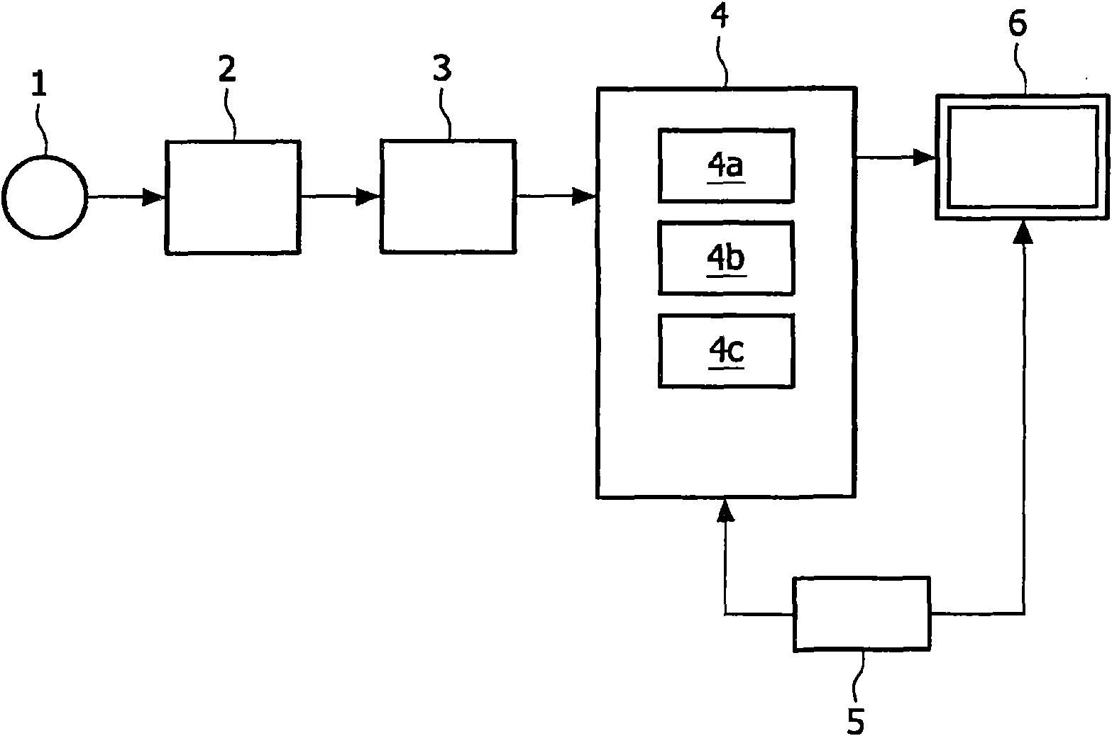
[0063] figure 1 A block diagram of a device according to the invention for imaging an object 1 is shown. Application of the data acquisition unit 2 on the object 1 or a part of the object 1 provides a three-dimensional (3D) data set. The unit 2 may be a unit arranged to perform magnetic resonance imaging (MRI), computed tomography (CT), ultrasound scanning, optical imaging or (3D) rotational angiographic x-rays on a subject.

[0064] Thus, the image data set is preferably a medical image data set, but the invention also relates to and is suitable for applications in conjunction with geological analysis, material analysis, architectural analysis and the like. Nevertheless, in the remainder of the description, the medical embodiment will be further explained, ie the subject 1 is a patient or a part of a patient. Specifically, the tubular structures studied in the present invention may be blood vessels, bones, airways, colons or vertebrae. In a specific embodiment, the blood ...


PUM


Abstract

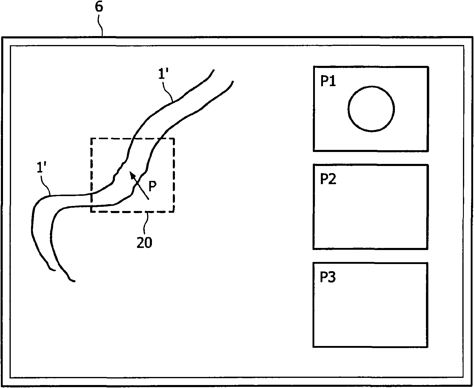
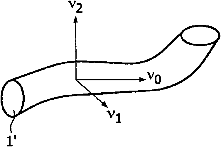
The present invention discloses a method for inspecting tubular-shaped structures (1 ) within a three-dimensional (3D) image data set, e.g. a vessel in a medical image. Initially, there is provided animage data set and performed a visualization of the image data set. Then, an inspection of the image data set is performed. During the inspection the user moves a pointer (P), e.g. via a computer mouse, and a processor performs a local segmentation around the pointer so as to determine a possible shape of a tubular shaped segmented object (1 ), e.g. a vessel, and the processor also makes a localanalysis of the segmented object. Thereafter, a screen displays a view (P1) of the segmented object (1 ), where the orientation of the first view is derived from the local analysis; the first view canfor example be cross-sectional or longitudinal views. The invention may be used directly on the raw image data in a great diversity of visualizations. No advanced application knowledge such as anatomical models, advanced acquisition protocol settings or global segmentation is needed. It is therefore a robust method that can be used over a wide range of image modalities and anatomies, which is essential in a vascular quantification package.

Description

technical field [0001] The present invention relates to a method for examining tubular structures within a three-dimensional (3D) image dataset, in particular a medical image dataset. The invention also relates to a corresponding imaging system and a corresponding computer program. Background technique [0002] The field of medical imaging, and specifically the field of multimodal 3D vessel analysis, is gaining more and more attention. Imaging tools provide advanced visualization, segmentation, inspection, and quantification of blood vessels for diagnosis across many patient populations. [0003] Most manufacturers of medical imaging workstations have tools to support vessel analysis, specifically 3D vessel quantification. [0004] Although the term 3D vessel quantification sounds like referring to a single application, it actually relates to a set of applications targeting different vessel structures using different acquisition methods, for which the need for the desired ...

Claims


Application Information

Patent Timeline
Patent Type & Authority Applications(China)
IPC IPC(8): G06T7/00
CPCG06T2207/30101G06T7/0012
Inventor J·J·松内曼斯R·J·E·哈比斯J·奥利万贝斯科斯
Owner KONINK PHILIPS ELECTRONICS NV
Learn More