
Spectral doppler ultrasound imaging device and method for automaticly controlling same

A technology of Doppler ultrasound and imaging equipment, applied in the directions of ultrasound/sound wave/infrasound image/data processing, sound wave re-radiation, ultrasound/sound wave/infrasound diagnosis, etc.

Inactive Publication Date: 2012-04-11
KONINK PHILIPS ELECTRONICS NV
View PDF3 Cites 10 Cited by
  • Summary
  • Abstract
  • Description
  • Claims
  • Application Information

AI Technical Summary

Problems solved by technology

However, since such techniques depend on acquiring image-live measurements, they have similar problems of switching between spectrum-live and image-live modes as described further above and may compromise the two

Method used


Image

  • Spectral doppler ultrasound imaging device and method for automaticly controlling same
  • Spectral doppler ultrasound imaging device and method for automaticly controlling same
  • Spectral doppler ultrasound imaging device and method for automaticly controlling same

Examples


Embodiment Construction

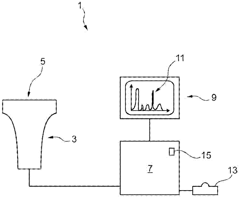
[0050] exist figure 1 , schematically depicts a spectral Doppler ultrasound imaging device 1 according to an embodiment of the present invention. The ultrasound transducer 3 comprises an ultrasound transceiver face 5 from which ultrasonic waves can be emitted into the patient's body, and the reflected echoes can then be detected by the ultrasound transceiver face 5 . The transducer 3 is connected to a control device 7 . The control device 7 can receive the detected echo signal from the transducer 3 and can display a corresponding ultrasound image on the display 9 based on the echo signal. In a first mode of operation, the control device 7 can control the transducer 3 to operate in an image-live measurement mode to provide a color or greyscale two-dimensional (2D) or three-dimensional (3D) ultrasound image to be displayed on the display 9 . In a second mode, the control device 7 can control the transducer 3 to operate in a spectrally live Doppler measurement mode and, for ex...


PUM


Abstract

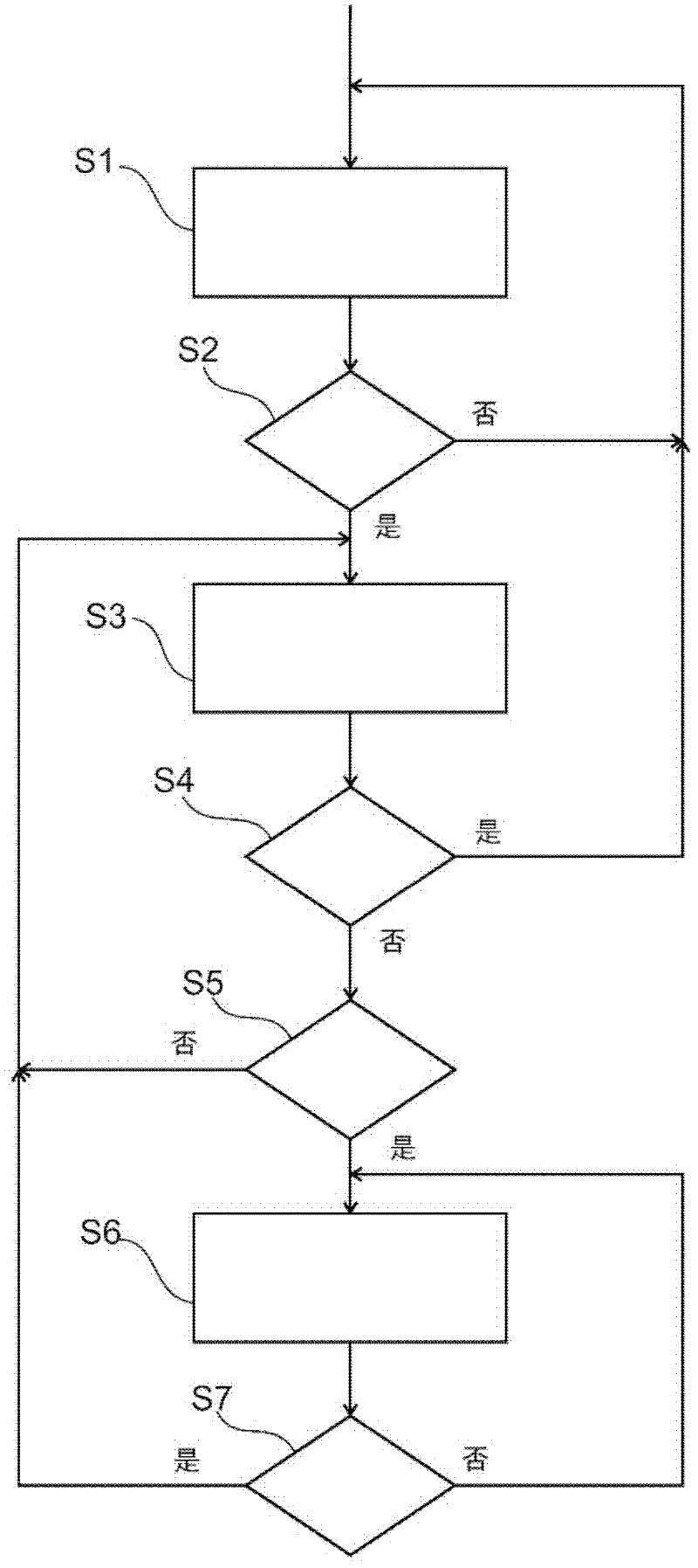
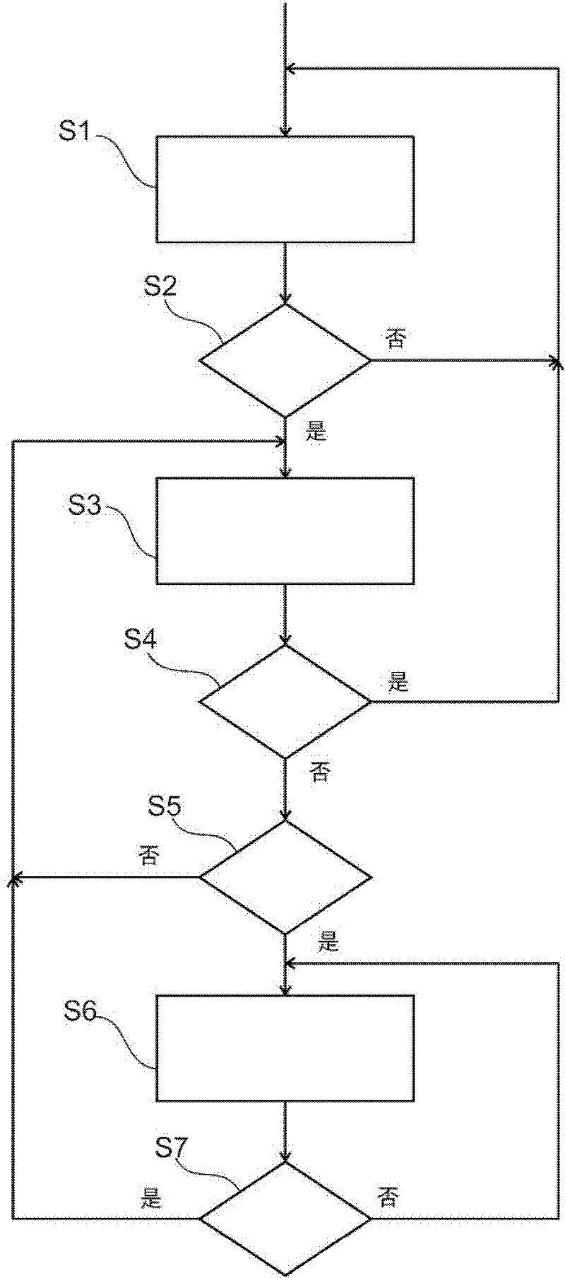
A method for controlling a spectral Doppler ultrasound imaging device adapted for operating both in a spectrum-live Doppler measurement mode (S3) and in an image-live measurement mode (S1, S6) is proposed. In accordance with the present invention, the method comprises automatically switching to the image-live measurement mode (S6) upon detection (S5) of a change of a sampled region. A change of a sampled region may be indicated by an operator by actuating a sampled region control device such as a trackball, angle knob or sample gate controller and, upon such actuation, the spectral Doppler ultrasound imaging device may automatically switch to the image-live measurement mode (S6) to allow for adapting or correcting of a position and / or orientation of the sampled region. After the sampled region has been positionally stable for a predetermined duration, the spectral Doppler ultrasound imaging device may be switched back (S7) to the spectrum-live measurement mode (S3). Thereby, unnecessary manual toggling between different measurement modes may be avoided and quantitative spectrum-live Doppler measurement (S3) is enabled being interrupted only when necessary, i.e. when a sampled region is changed.

Description

technical field [0001] The invention relates to a method for controlling a spectral Doppler imaging device adapted to operate both in a spectral live Doppler measurement mode and in an image live measurement mode. Furthermore, the invention relates to such a spectral Doppler ultrasound imaging device adapted to carry out the method of the invention, a computer program product for correspondingly controlling the spectral Doppler ultrasound imaging device when executed on a computer and having stored thereon A computer-readable medium of such a computer program product. Background technique [0002] Ultrasound imaging devices have been developed to qualitatively and quantitatively image both static structures and dynamic motion within a sampled area. Therein, ultrasonic waves are emitted toward the sampling area, and echoes reflected in the sampling area are detected. A sampling region, sometimes referred to as a region of interest, may be a part of the patient's body. The ...

Claims


Application Information

Patent Timeline
Patent Type & Authority Applications(China)
IPC IPC(8): G01S7/52G01S15/89A61B8/08
CPCA61B8/488A61B8/13G01S7/52063G01S7/52084G01S15/8979A61B8/06G01S7/52066
Inventor D·克拉克
Owner KONINK PHILIPS ELECTRONICS NV
Learn More