Ultrasonic diagnostic apparatus and region-of-interest

A technology of concerned area and diagnostic device, which is applied in the directions of acoustic wave diagnosis, infrasonic wave diagnosis, ultrasonic/sonic wave/infrasonic wave diagnosis, etc., to achieve the effect of improving accuracy

Inactive Publication Date: 2012-05-23
HITACHI MEDICAL CORP
View PDF5 Cites 7 Cited by
  • Summary
  • Abstract
  • Description
  • Claims
  • Application Information

AI Technical Summary

Problems solved by technology

[0008] However, in Patent Document 1, only a process of setting an ROI on the anterior wall and the posterior wall of a blood vessel is proposed, because there is no aspect of setting an ROI by associating and referring to a plurality of contour candidate points related to IMT measurement in a blood vessel. Considering this, there are unresolved issues regarding the accuracy of ROI setting related to IMT measurement

Method used


Image

  • Ultrasonic diagnostic apparatus and region-of-interest
  • Ultrasonic diagnostic apparatus and region-of-interest
  • Ultrasonic diagnostic apparatus and region-of-interest

Examples


Embodiment 1

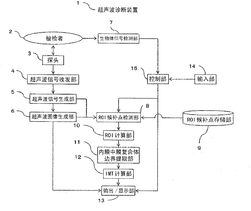
[0028] figure 1 It is a block diagram showing the outline of the ultrasonic diagnostic apparatus of the first embodiment of the present invention.

[0029] In the first embodiment, the function of "transmitting and transmitting ultrasound to the part including the carotid artery of the subject to take an ultrasound image" is composed of the ultrasound probe 3, the ultrasound signal transmitting and receiving unit 4, the ultrasound signal generating unit 5, and the ultrasound image generating unit 6 to bear.

[0030] In addition, a function of "scanning the ultrasound image by the region of interest setting unit and setting the region of interest including the intima-media complex on the ultrasound image based on the concentration of the carotid artery contour candidate points" is performed The ROI candidate point detection unit 8, the ROI candidate point storage unit 9 and the ROI calculation unit 10 are responsible.

[0031] In addition, the function of “measurement of the thicknes...

Embodiment 2

[0115] In the second embodiment, a case where the number of ROIs is plural is exemplified and described. Since the configuration and operation of the ultrasonic diagnostic apparatus 1 are the same as those of the first embodiment, the description is omitted, and only the parts that are different from the first embodiment will be described.

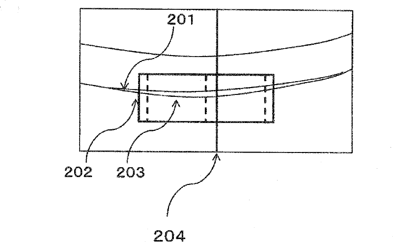
[0116] use Image 6 The calculation process of the position and size of the ROI will be described.

[0117] Image 6 It is a diagram for explaining the principle of ROI setting in Embodiment 2 of the present invention.

[0118] First, the candidate point data 603 is drawn on the ultrasound image of the carotid artery displayed in the image display area 502 on which the screen is drawn. Then, a plurality of candidate point data 603 are obtained in the same order, and the plurality of candidate point data 603 are similarly drawn on the ultrasound image. The coordinate points of the drawn candidate point data 603 on the ultrasound image are stored...

Embodiment 3

[0132] In the third embodiment, an example will be described in which the ROI setting performed on the blood vessel wall of the (one) carotid artery near the probe is reflected in the ROI setting of the blood vessel wall (the other) farther from the probe.

[0133] Since the configuration and operation of the ultrasonic diagnostic apparatus 1 are the same as those of the first embodiment, the description is omitted, and only the differences from the first embodiment will be described.

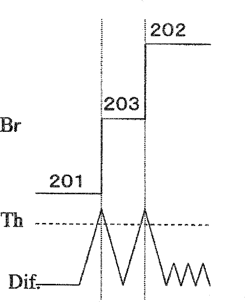
[0134] use Figure 7 The calculation process of the position and size of the ROI in this embodiment will be described.

[0135] Figure 7 It is a diagram explaining the principle of ROI setting in the third embodiment of the present invention.

[0136] First, on the side of the carotid artery, the outer wall part on the lower side of the figure and image 3 Perform data processing in the same way.

[0137] Next, data processing is performed on the outer wall portion on the other side of the carotid arter...


PUM


Abstract

Disclose is an ultrasonic diagnostic apparatus which transmits / receives an ultrasonic wave to / from a region including a carotid artery of an object, images an ultrasonic image, and comprises a thickness measurement unit configured to measure the thickness of an intima-media complex from the ultrasonic image, the ultrasonic diagnostic apparatus being provided with a region-of-interest setting unit configured to scan the ultrasonic image and set a region of interest including the intima-media complex on the ultrasonic image on the basis of the degree of concentration of contour slate points of the carotid artery, wherein the thickness measurement unit measures the thickness of the intima-media complex on the basis of boundaries within the set region of interest.

Description

Technical field [0001] The present invention relates to setting a region of interest (Region Of Interest: ROI) for measuring the thickness of the intima media complex (Intima Media Thickness: IMT) in a captured ultrasound image of the carotid artery of a subject and Ultrasonic diagnostic device that measures IMT value and its area of ​​interest setting method. Background technique [0002] It is known that IMT measurement is relatively effective for detecting arteriosclerosis of the subject. In IMT measurement, the following process is required: a medical practitioner such as a physician or a clinical examination technician (referred to as an "examiner") places an ultrasound probe against the examinee’s carotid artery to obtain an ultrasound image and set it on the ultrasound image ROI. [0003] The examiner can set the ROI even manually. However, it is impossible to perform effective image diagnosis only by the examiner performing the ROI setting operation related to IMT measur...

Claims


Application Information

Patent Timeline
Patent Type & Authority Applications(China)
IPC IPC(8): A61B8/00
CPCG06T7/602A61B8/06G06T7/0012G06T2207/10132A61B8/5223A61B8/469A61B8/0891G06T2207/30101A61B8/02G06T7/62G16H50/30
Inventor 长野智章
Owner HITACHI MEDICAL CORP
Why Eureka
Try Eureka