Diagnosis assisting system, diagnosis assisting program, and diagnosis assisting method

A diagnostic aid, vascular technology used in diagnosis, diagnostic recording/measurement, computed tomography, etc.

Inactive Publication Date: 2012-07-04
FUJIFILM CORP
View PDF7 Cites 8 Cited by
  • Summary
  • Abstract
  • Description
  • Claims
  • Application Information

AI Technical Summary

Problems solved by technology

However, current research suggests health risks even at low stenosis rates

Method used


Image

  • Diagnosis assisting system, diagnosis assisting program, and diagnosis assisting method
  • Diagnosis assisting system, diagnosis assisting program, and diagnosis assisting method
  • Diagnosis assisting system, diagnosis assisting program, and diagnosis assisting method

Examples


Embodiment Construction

[0034] Hereinafter, embodiments of the diagnostic assistance device, diagnostic assistance program, and diagnostic assistance method of the present invention will be described based on the accompanying drawings.

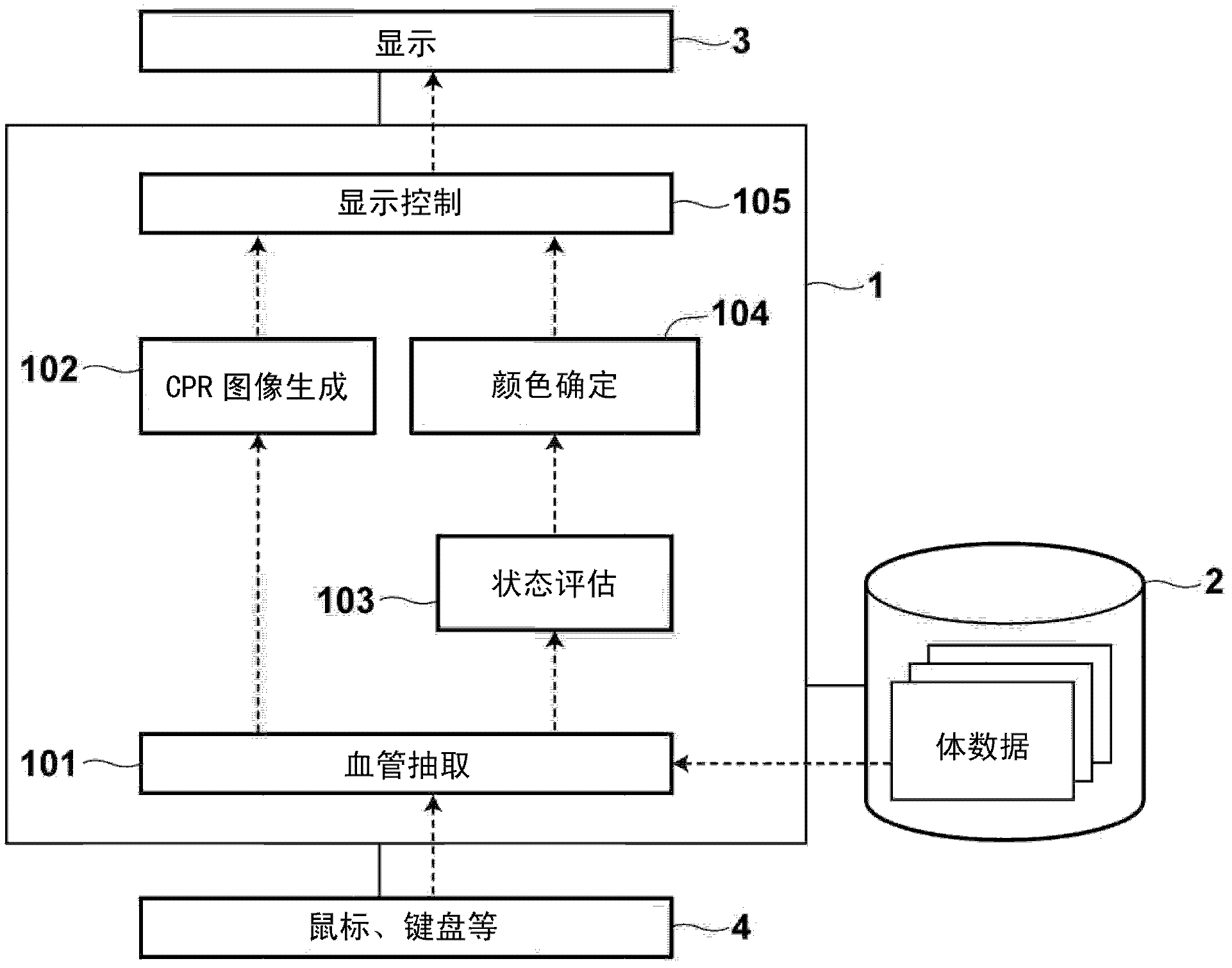
[0035] figure 1 A schematic configuration of a diagnostic assistance apparatus 1 obtained by installing a diagnostic assistance program in a workstation to be used by a physician is illustrated. The diagnostic aid 1 is equipped with a processor and memory (not shown) as standard workstation components. The diagnostic aid 1 is also equipped with storage 2 such as HDD (Hard Disk Drive) and SSD (Solid State Drive). Furthermore, a display 3 and input devices 4 such as a keyboard and a mouse are connected to the diagnostic aid 1 .

[0036] When the diagnostic support program is installed in the workstation, the diagnostic support program and data referred to by the diagnostic support program (conversion table, etc. which will be described later) are stored in the memory...


PUM


Abstract

A CPR image (14) showing at least partial region of a blood vessel is generated from volume data obtained by image capturing and is arranged in a predetermined region of a display screen (11C). Outside this region, band-shaped condition presentation regions (13, 15) are arranged so as to be parallel with the blood vessel shown by the CPR image. A narrow ratio and the indication value of, for example, the presence and absence of plaque are calculated by a volume data analysis. The calculated indication value is replaced with a color (RGB value) with reference to a conversion table. The condition presentation regions (13, 15) are color-coded according to the indication value and displayed. This makes it possible to, on a blood vessel diagnosis screen, extensively and sufficiently observe the conditions of a blood vessel inner wall and simultaneously to intuitively understand the narrow ratio and the like in each region of the blood vessel.

Description

technical field [0001] The present invention relates to a diagnosis assistance system, a diagnosis assistance computer program and a diagnosis assistance method for assisting diagnosis of blood vessels by analyzing and visualizing three-dimensional image data. Background technique [0002] Early detection of stenosis is very important to prevent ischemic diseases such as myocardial infarction and cerebral infarction. For this reason, recently, a diagnostic aid equipped with a blood vessel analysis function and blood vessel analysis software has become available. For example, Patent Document 1 discloses a blood vessel stenosis rate analysis system that detects a stenosis site of a blood vessel by analyzing volume data obtained by CT imaging and MRI imaging, and calculates the stenosis rate and region stenosis rate of a blood vessel. This system displays a three-dimensional image of a blood vessel generated by surface rendering or volume rendering with colors added based on t...

Claims


Application Information

Patent Timeline
Patent Type & Authority Applications(China)
IPC IPC(8): A61B6/03A61B5/00A61B5/055
CPCG06T19/20G06T2207/10072G06T15/08G06T2215/06G06T2219/2012G06F19/30G06T11/206G06T2200/24G06T2207/30101G06T2207/30048G06T19/003G06T2207/30172G06T7/0012
Inventor 平野雅春樱木太桝本润
Owner FUJIFILM CORP
Why Eureka
Try Eureka