Orthopedic fixation with imagery analysis

An orthopedic and fixator technology, applied in external fixators, fixators, stereotaxic surgical instruments, etc., can solve problems such as improper alignment of bone segments and inaccurate imaging processes

Active Publication Date: 2013-01-16
SYNTHES GMBH
View PDF3 Cites 21 Cited by
  • Summary
  • Abstract
  • Description
  • Claims
  • Application Information

AI Technical Summary

Problems solved by technology
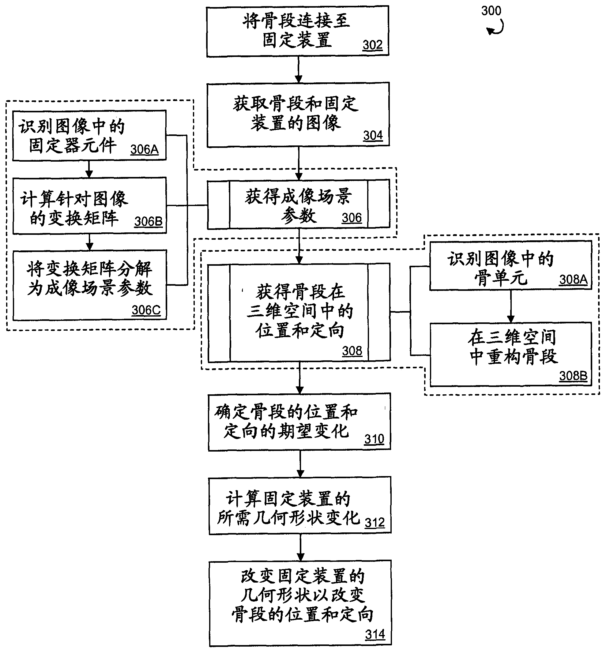
[0004] However, the ability to obtain orthoradiographic images of the fracture site can be limited by factors beyond the surgeon's control, such as the maneuverability of the imaging device, the anatomical location of the fracture or deformity, and/or the ability to locate the severed limb for use. Patient Pain During Orthogonal Imaging
Limiting factors su...

Method used


Image

  • Orthopedic fixation with imagery analysis
  • Orthopedic fixation with imagery analysis
  • Orthopedic fixation with imagery analysis

Examples


Embodiment Construction

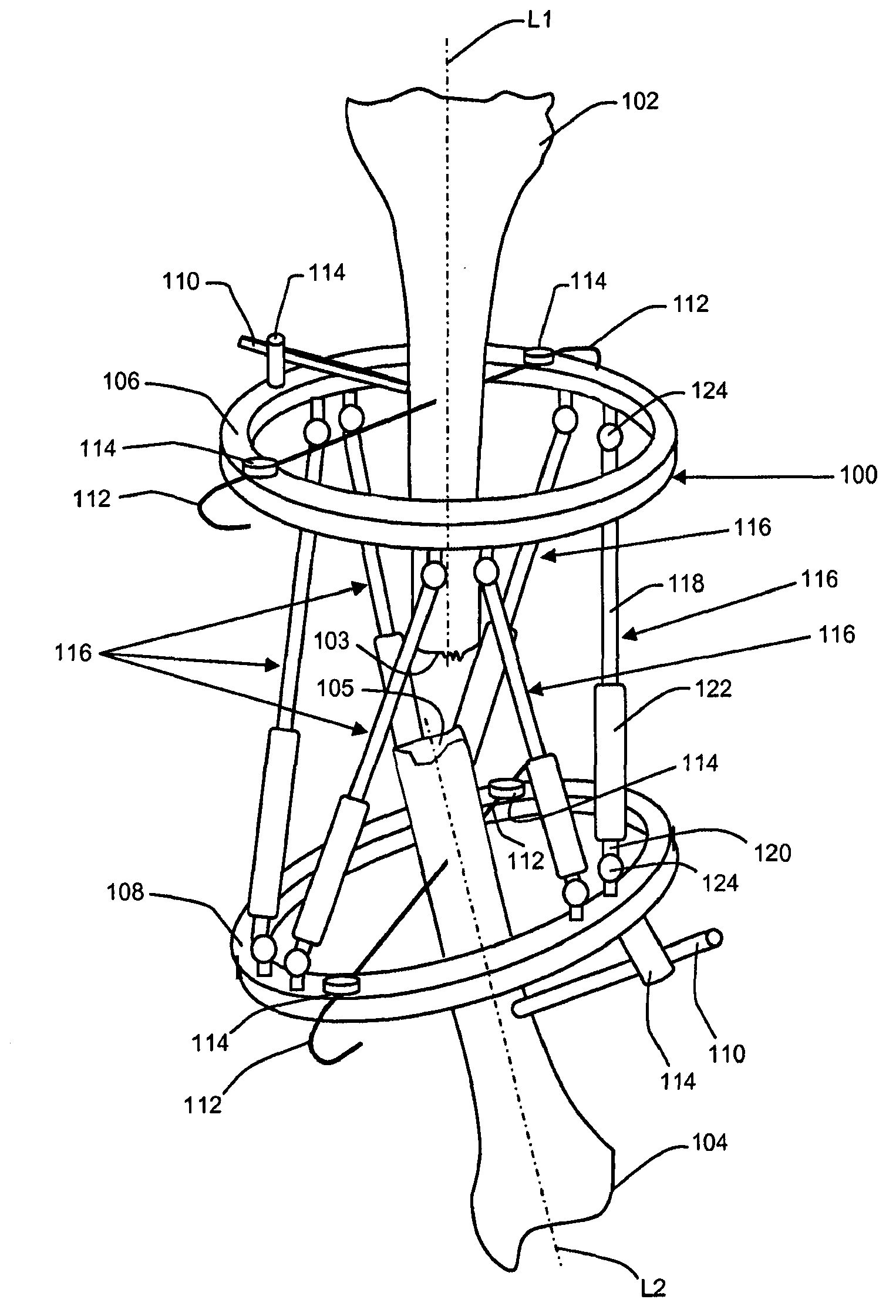
[0011] For convenience, the same or equivalent elements in the various embodiments illustrated in the figures have been labeled with the same reference numerals. Certain terms are used in the following description for convenience only and not for limitation. The words "right", "left", "top" and "bottom" refer to directions in the drawings to which reference is made. The words "inwardly," "inwardly," "outwardly," and "outwardly" refer to directions toward and away from, respectively, the geometric center of the device and its referenced parts. Terms intended to be non-limiting include the words listed above, their derivatives and words of similar import.

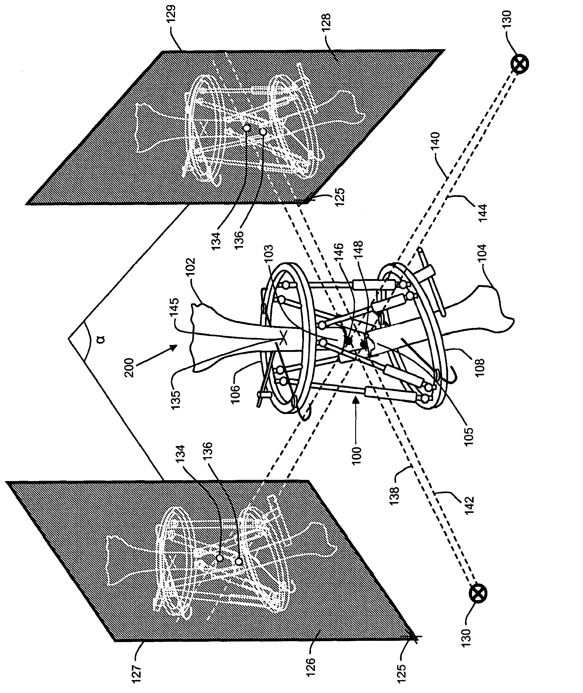
[0012] First refer to figure 1 , body tissue (eg, first bone segment 102 and second bone segment 104 ) may be aligned and / or oriented to promote union or other healing between the body tissues. Alignment and / or orientation of body tissue can be achieved by coupling the body tissue to an adjustable fixation device (eg, orth...


PUM


Abstract

Methods of orthopedic fixation and imagery analysis are provided. Images of first and second bone segments attached to a fixation apparatus are captured. Fixator elements identified in the images can be used to obtain imaging scene parameters. Bone elements identified in the images can be used with the imaging scene parameters to reconstruct a three dimensional representation of positions and/or orientations of the first and second bone segments with respect to the fixation apparatus.

Description

[0001] Cross References to Related Applications [0002] This patent application claims priority from UK patent application serial number GB1008281.6 filed 19 May 2010, the entire content of which application is hereby incorporated by reference. Background technique [0003] Techniques for treating fractures and / or bone deformities may include the use of external fixators (eg, fixation frames) that are surgically mounted to bone segments on opposite sides of the fracture site. Paired radiographic images of the fixator and bone segment at the fracture site are taken. In general, radiographic images must be orthogonal or perpendicular relative to each other and aligned with the patient's anatomical axis. Data from the images is then manipulated using orthogonal projection techniques to construct a three-dimensional representation of the fixture and bone segments that can be used in developing a treatment plan, and which may include, for example, Bone segments realigned. [00...

Claims


Application Information

Patent Timeline
IPC IPC(8): A61B17/62A61B17/66A61B19/00G16Z99/00
CPCA61B2019/5295A61B2019/5466A61B17/62A61B17/66A61B19/203A61B19/50A61B2090/367A61B90/14A61B2090/3966G16H30/40G16H20/40A61B34/10G16Z99/00
Inventor A·尼昆诺瓦斯
Owner SYNTHES GMBH
Why Eureka
Try Eureka