Virtual X-ray imaging method and virtual X-ray imaging system for human body bone joint

An imaging method and imaging system technology, applied in the field of medical imaging, can solve the problems of inconvenient real-time adjustment and observation, complicated operation, and high cost

Inactive Publication Date: 2013-08-14
THE FIRST AFFILIATED HOSPITAL OF THIRD MILITARY MEDICAL UNIVERSITY OF PLA
View PDF4 Cites 5 Cited by
  • Summary
  • Abstract
  • Description
  • Claims
  • Application Information

AI Technical Summary

Problems solved by technology

Observing and understanding these effects often requires repeated X-rays of the human body, which is not only costly, complicated to operate, and increases radiation intake, but also cannot be easily adjusted and observed in real time, making it difficult to implement in reality

Method used


Image

  • Virtual X-ray imaging method and virtual X-ray imaging system for human body bone joint
  • Virtual X-ray imaging method and virtual X-ray imaging system for human body bone joint
  • Virtual X-ray imaging method and virtual X-ray imaging system for human body bone joint

Examples


Embodiment Construction

[0043] The preferred embodiments of the present invention will be described in detail below in conjunction with the accompanying drawings; it should be understood that the preferred embodiments are only for illustrating the present invention, rather than limiting the protection scope of the present invention.

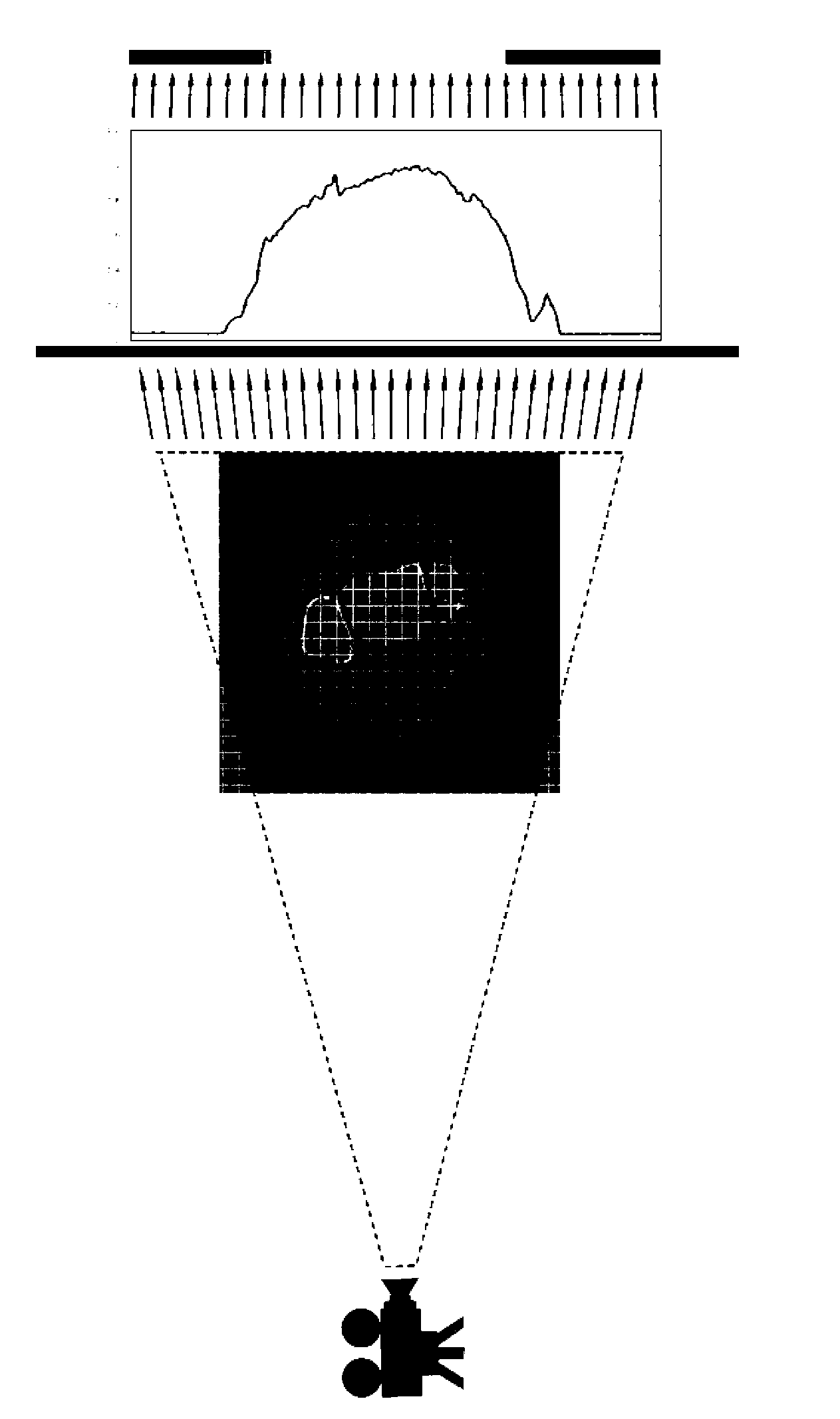
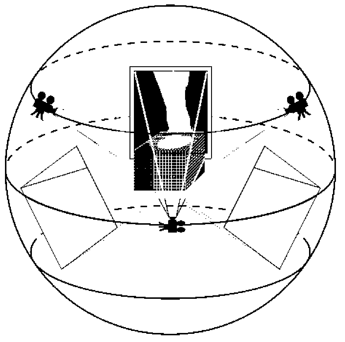
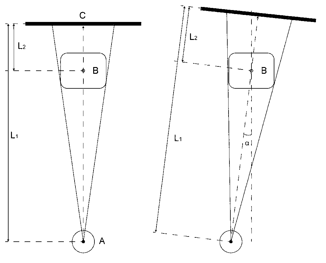
[0044] figure 1 is a schematic diagram of the virtual imaging plane, figure 2 is a stereoscopic schematic diagram of virtual imaging, image 3 In order to simulate different projection angles, focal lengths and object distances, in the figure, L1 represents the object distance, L2 represents the focal distance, Figure 4 For selective virtual imaging, Figure 5 It is a flowchart of a virtual X-ray imaging method for human bone joints, as shown in the figure: The virtual X-ray imaging method for human bone joints provided by the present invention includes the following steps:

[0045] The human bone joint virtual X-ray imaging method provided by the present invention...


PUM


Abstract

The invention discloses a virtual X-ray imaging method and a virtual X-ray imaging system for a human body bone joint. The method comprises the steps that computed tomography (CT) is conducted on a targeted limb; the limb is converted into a data set formed by coordinates of a volume unit and an X-ray absorption rate; a course that a ray reaches an imaging plain through the limb is simulated by a numerical algorithm; visualization treatment is conducted on a numerical result; a gray level image that can be identified by a human eye is generated; and the result is outputted. For orthopedic and radio-diagnosis doctors, X-ray images generated by projection to the limb from any angles can be generated; the orthopedic and radio-diagnosis doctors can be assisted in learning X-ray anatomy, and establishing cognition connection between a three-dimensional structure and a two-dimensional image; for a radiographer, influences of different shooting parameters and a body position of an examined person on final imaging can be simulated; the method and the system contributes to learning a radiography technology of the radiographer; and for a special disease, the method and the system can assist in screening the optimal X-ray radiography body position and the angle for clinical reference.

Description

technical field [0001] The invention relates to the field of medical imaging, in particular to a method and system for virtual X-ray imaging of human bone joints. Background technique [0002] X-ray film is an important method in the imaging diagnosis of bone and joint diseases. Although the development of medical imaging technologies such as computed tomography (CT) and magnetic resonance imaging (MRI) has provided new ways for orthopedic diagnosis, the information provided by traditional plain X-ray images is still irreplaceable. In addition, X-ray plain film imaging has the advantages of high image clarity, low radiation dose, low price, simple and fast, and can be examined under load, so it is still an indispensable means of clinical examination. [0003] X-ray plain film images are superimposed and projected on a two-dimensional plane in the perspective of the tissue structure in the direction of ray penetration. The images of different structures overlap each other, w...

Claims


Application Information

Patent Timeline
Patent Type & Authority Applications(China)
IPC IPC(8): A61B6/03
Inventor 尹力杨柳
Owner THE FIRST AFFILIATED HOSPITAL OF THIRD MILITARY MEDICAL UNIVERSITY OF PLA
Why Eureka
Try Eureka