Method and device for locating cervical vertebral body axis and related tissues in MRI image

A technology of vertebral body axis and positioning method, which is applied in medical science, sensors, diagnostic recording/measurement, etc., and can solve problems such as matching failure, not very effective, and time-consuming

Inactive Publication Date: 2015-11-25
SHENZHEN MINDRAY BIO MEDICAL ELECTRONICS CO LTD
View PDF5 Cites 2 Cited by
  • Summary
  • Abstract
  • Description
  • Claims
  • Application Information

AI Technical Summary

Problems solved by technology

However, this segmentation method is often time-consuming and not very effective, because the gray value of the intervertebral disc will change in MRI images with different weights. T1 inversion recovery) weight image is white
In addition, there are some methods that use deformable model matching to extract the vertebral body and then locate the intervertebral disc. The problem with this method is that the number of visible vertebral bodies in the spine image is not necessarily the same. If there are too many or too few visible vertebral bodies, the matching will fail.
Some segmentation methods require doctors to perform certain interactive operations, such as selecting feature points, which will reduce the efficiency of inspection

Method used


Image

  • Method and device for locating cervical vertebral body axis and related tissues in MRI image
  • Method and device for locating cervical vertebral body axis and related tissues in MRI image
  • Method and device for locating cervical vertebral body axis and related tissues in MRI image

Examples


Embodiment Construction

[0024] The present invention will be further described in detail below through specific embodiments in conjunction with the accompanying drawings.

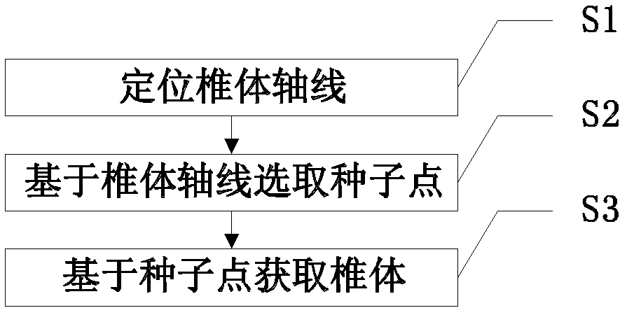
[0025] In each embodiment of the present invention, the identification and positioning of the cervical vertebral body and the cervical intervertebral disc usually involves first positioning the "vertebral body axis" and the "trachea line". concept. The said vertebral body axis in each embodiment of the present invention is not strictly limited to the line passing through the center of the vertebral body, as long as it can pass through all the vertebral bodies longitudinally, so the vertebral body axis defined in the various embodiments of the present invention is Some errors are allowed. Since the trachea region is in the shape of a long and narrow band in the entire magnetic resonance image, the detected trachea region is in the same line, so the trachea region is described here as a trachea line.

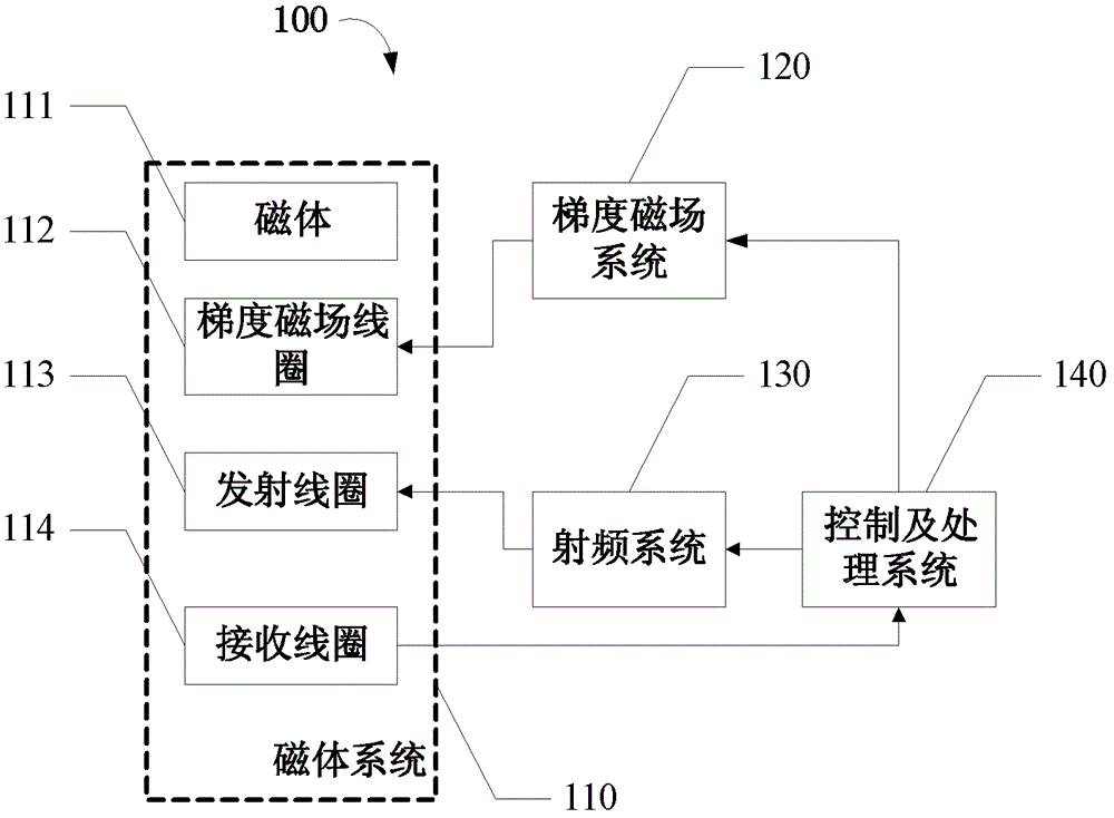
[0026] figure 1 Shown is th...


PUM


Abstract

The invention discloses a positioning method and a positioning device for cervical vertebral body axial lines and relevant tissues. The cervical vertebral body axial line is fast positioned through positioning the trachea line, in addition, the vertebral body is extracted in a self-adaptive way by utilizing the characteristics that the gray levels in the vertebral body are approximately uniform, in addition, the vertebral body has the obvious boundary with the intervertebral disc, and the shape and the size of the vertebral body are basically fixed. The extraction process is not influenced by the image weight, and the middle line position and the angle of each intervertebral disc are calculated according to the extracted vertebral body, so the scanning of the cervical intervertebral disc in an MRI (magnetic resonance imaging) system is effectively and automatically positioned. The invention also discloses a magnetic resonance system.

Description

technical field [0001] The invention relates to medical imaging equipment, in particular to a method and device for automatic positioning of cervical intervertebral discs by using magnetic resonance images, and a magnetic resonance imaging system. Background technique [0002] Magnetic Resonance Imaging (MRI, Magnetic Resonance Imaging) has been widely used clinically due to the advantages of non-invasive and multi-parameter imaging, especially its ability of arbitrary tomographic imaging, which can directly observe and analyze tissue structure and its lesions from different angles, so It has outstanding advantages in the serial examination of the spine. A typical MRI examination process of cervical intervertebral disc often requires physicians to manually place each group of scanning lines on the intervertebral disc with disease on the sagittal plane positioning image. In order to ensure that the thread group passes through the center of the intervertebral disc, it is nece...

Claims


Application Information

Patent Timeline
Patent Type & Authority Patents(China)
IPC IPC(8): A61B5/055
Inventor 云天梁邓晓云
Owner SHENZHEN MINDRAY BIO MEDICAL ELECTRONICS CO LTD
Why Eureka
Try Eureka