Cardiac real-time cine imaging image processing method and system

An image processing and film technology, applied in the field of image processing, can solve the problems of complicated and time-consuming pictures, and achieve the effects of saving time, improving processing efficiency, and simple operation

Active Publication Date: 2015-07-29
SHANGHAI UNITED IMAGING HEALTHCARE
View PDF4 Cites 2 Cited by
  • Summary
  • Abstract
  • Description
  • Claims
  • Application Information

AI Technical Summary

Problems solved by technology

[0005] Based on this, it is necessary to solve the cumbersome and time-consuming problem of manual selection of end-diastolic and end-systolic images at the end of expiration, and provide a simple and time-saving cardiac real-time cine imaging image processing method

Method used


Image

  • Cardiac real-time cine imaging image processing method and system
  • Cardiac real-time cine imaging image processing method and system
  • Cardiac real-time cine imaging image processing method and system

Examples


Embodiment Construction

[0039] The technical solution of the real-time cardiac cine imaging image processing method and system will be described in detail below in conjunction with specific embodiments and accompanying drawings, so as to make it more clear.

[0040] Such as figure 1 Shown is a kind of cardiac real-time cine imaging image processing method in an embodiment, comprising:

[0041] Step S102, the step of locating the right ventricle, setting a reference point from the center of the left ventricle selected from the real-time cine imaging image of the heart, and locating the right ventricle according to the reference point.

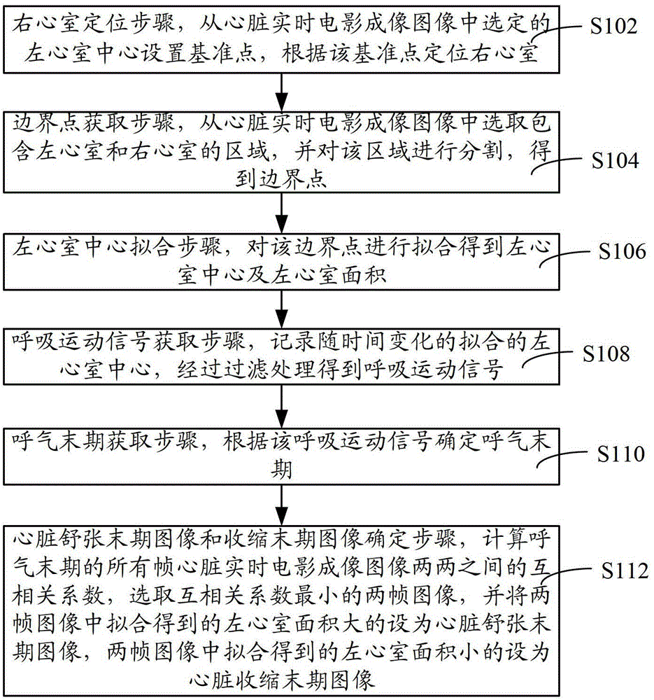
[0042] First, manually locate the left ventricle from cardiac live cine images and set a fiducial point at the center of the left ventricle. Such as figure 2 Shown is a schematic diagram of setting the reference point position, figure 2 The center of the fan-shaped area is the reference point.

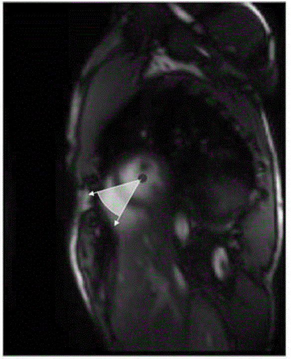
[0043] The step of locating the right ventricle according to the ref...


PUM


Abstract

The invention relates to a method and system for processing a heart real-time film imaged picture. The method comprises the steps that the center of the left ventricle is selected from the heart real-time film imaged picture so that a datum point can be arranged and the right ventricle can be located according to the datum point; a region is selected from the heart real-time film imaged picture and is divided so that a boundary point can be obtained; fitting is conducted on the boundary point so that the center of the left ventricle and the area of the left ventricle can be obtained; the fitted center, changing along with the time, of the left ventricle is recorded so that a breathing movement signal can be obtained and an end expiration period can be determined; a cross correlation coefficient between every two frame pictures in the expiration rear period is calculated, the two frames of pictures with the smallest correlation coefficient are selected, the picture of the left ventricle with the large area obtained through fitting serves as an end diastolic picture and the picture of the left ventricle with the small area obtained through fitting serves as an end-systole picture. According to the method and system for processing the heart real-time film imaged picture, operation is simple, time is saved, and the cardiac function can be analyzed conveniently, rapidly and reliably.

Description

technical field [0001] The invention relates to the field of image processing, in particular to an image processing method and system for cardiac real-time cine imaging. Background technique [0002] Cardiac magnetic resonance cine imaging is a commonly used imaging method for measuring cardiac function in clinical practice. Generally, part of the K-space data is collected within one cardiac cycle. Since the complete k-space data is completed over multiple cardiac cycles, the method is sensitive to respiratory motion. In order to eliminate the influence of breathing movement on the imaging quality, the subjects need to hold their breath repeatedly during the scanning process. This requirement is difficult to achieve in patients with severe heart disease, respiratory disease and infants, so this method has greater clinical limitations. [0003] Cardiac magnetic resonance real-time cine imaging can complete the acquisition of all K-space data within one cardiac cycle by appr...

Claims


Application Information

Patent Timeline
Patent Type & Authority Patents(China)
IPC IPC(8): A61B5/055
Inventor 吴垠江克钟耀祖刘新郑海荣
Owner SHANGHAI UNITED IMAGING HEALTHCARE
Why Eureka
Try Eureka