X-ray diagnostic system

A diagnostic system and X-ray technology, applied in computerized tomography scanners, echo tomography, etc., can solve the problems of inability and constant correction, and achieve the effect of improving work efficiency

Inactive Publication Date: 2014-02-12
NANJING JOOZ INFORMATION SCI & TECH
View PDF0 Cites 0 Cited by
  • Summary
  • Abstract
  • Description
  • Claims
  • Application Information

AI Technical Summary

Problems solved by technology

[0009] The technical problem to be solved by the present invention is to provide a method that can correct the above-mentioned defects such as the correction amount of the CT perfusion image and the X-ray perfusion image of the X-ray diagnostic device in the prior art is

Method used


Image

  • X-ray diagnostic system
  • X-ray diagnostic system

Examples


Embodiment Construction

[0034] In order to make the object, technical solution and advantages of the present invention clearer, the present invention will be further described in detail below in conjunction with the accompanying drawings and embodiments. It should be understood that the specific embodiments described here are only used to explain the present invention, not to limit the present invention.

[0035] Preferred embodiments of the present invention are as follows:

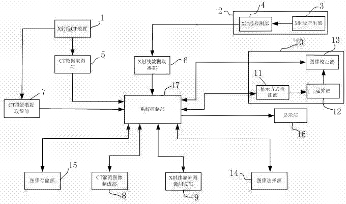
[0036] An X-ray diagnostic system such as figure 1 As shown, it includes an X-ray CT apparatus 1, an X-ray diagnostic apparatus 2, a data acquisition unit, a CT perfusion image creation unit 8, an X-ray perfusion image creation unit 9, a correction unit 10, an image selection unit 14, and an image storage unit 15 , a display unit 16 and a system control unit 17, wherein the data acquisition unit includes a CT data acquisition unit 5, an X-ray data acquisition unit 6 and a CT projection data acquisition unit 7, and the correcti...


PUM


Abstract

The invention discloses an X-ray diagnostic system which comprises an X-ray CT (computed tomography) device, an X-ray diagnostic device, a data obtaining part, a CT perfusion picture producing part, an X-ray perfusion picture producing part, a correction part, a picture selection part, a picture storage part, a display part and a system control part, wherein the correction part comprises a picture correction part, a display mode detection part and an operation part. By adopting the X-ray diagnostic system, a CT perfusion picture can be rectified with a correcting value changed at any time, so that the display mode of the CT perfusion picture and that of an X-ray perfusion picture can be coincident; in addition, the work efficiency of a doctor comparing various perfusion pictures is improved.

Description

[0001] technical field [0002] The invention relates to a medical diagnosis system, in particular to an X-ray diagnosis system. [0003] Background technique [0004] Currently, in the examination of ischemic heart disease in the field of tracheal medicine, X-ray CT (Computed Tomography) equipment is generally used for diagnosis first, and when it is diagnosed that treatment is required, X-ray diagnostic equipment is generally used for treatment . And, during the treatment, by comparing the CT myocardial perfusion image made from the CT image taken by the X-ray CT device with the X-ray myocardial perfusion image made from the X-ray projection image produced by the X-ray diagnostic device, the treatment can be concluded. Program. [0005] Myocardial perfusion images can represent blood flow dynamics in the myocardium. Myocardial perfusion images are generated by generating time-density curves related to predetermined index values ​​representing blood dynamics from imag...

Claims


Application Information

Patent Timeline
IPC IPC(8): A61B6/03
Inventor 周新法
Owner NANJING JOOZ INFORMATION SCI & TECH
Why Eureka
Try Eureka