Polypeptide for inhibiting cell proliferation and inducing apoptosis and preparation method and use thereof

A technique for inhibiting cell and apoptosis, applied in the field of polypeptides that inhibit cell proliferation and induce apoptosis and its preparation, achieve great clinical application value, induce apoptosis, and have obvious effects

Active Publication Date: 2014-03-05
TONGJI HOSPITAL ATTACHED TO TONGJI MEDICAL COLLEGE HUAZHONG SCI TECH
View PDF1 Cites 2 Cited by
  • Summary
  • Abstract
  • Description
  • Claims
  • Application Information

AI Technical Summary

Benefits of technology

This patented technology allows researchers to study how magnetic particles (Magnetic Resonance) help block growth factors called cell signal transducers from affecting neovasculature formation during medical procedures like surgery. These agents were found to cause inflammations within arteries when injected into patients' veins without causing damage. By administering these medicines through injection, this method could potentially improve treatments for various types of cancer by reducing tumor size and promoting better outcomes postoperatively.

Problems solved by technology

This patents describes how certain compounds are able to stop or slow up growth of nerve fibers involved with vasculature receptors like endothecium Schwann Cell Stroke Pathway(ESCRP)/mitogen activator nuclear factor 1α complexes (MASCs)). However, these substances have been shown to cause drug resistance leading to decreased effectiveness against this mechanism. To address this problem, we propose modifying existing therapeutic agents based on their ability to enhance the activity of targets associated with angiotensin converting enzyme (ACE)-mediating loop proteins including ASCLS5, KIN-1, LSP, CARDI-5B, CELL_HAND_VERF11a, MEISENICISTS, SHDN-314, SEQ ID NO:1, NITOX, TGR20, FIG3. Overall, there is an urgent technical issue addressed by this patented paper relates to finding ways to improve the effects of chemotherapy treatments without causing side effects due to therapies themselves.

Method used


Image

  • Polypeptide for inhibiting cell proliferation and inducing apoptosis and preparation method and use thereof
  • Polypeptide for inhibiting cell proliferation and inducing apoptosis and preparation method and use thereof
  • Polypeptide for inhibiting cell proliferation and inducing apoptosis and preparation method and use thereof

Examples


experiment example 1

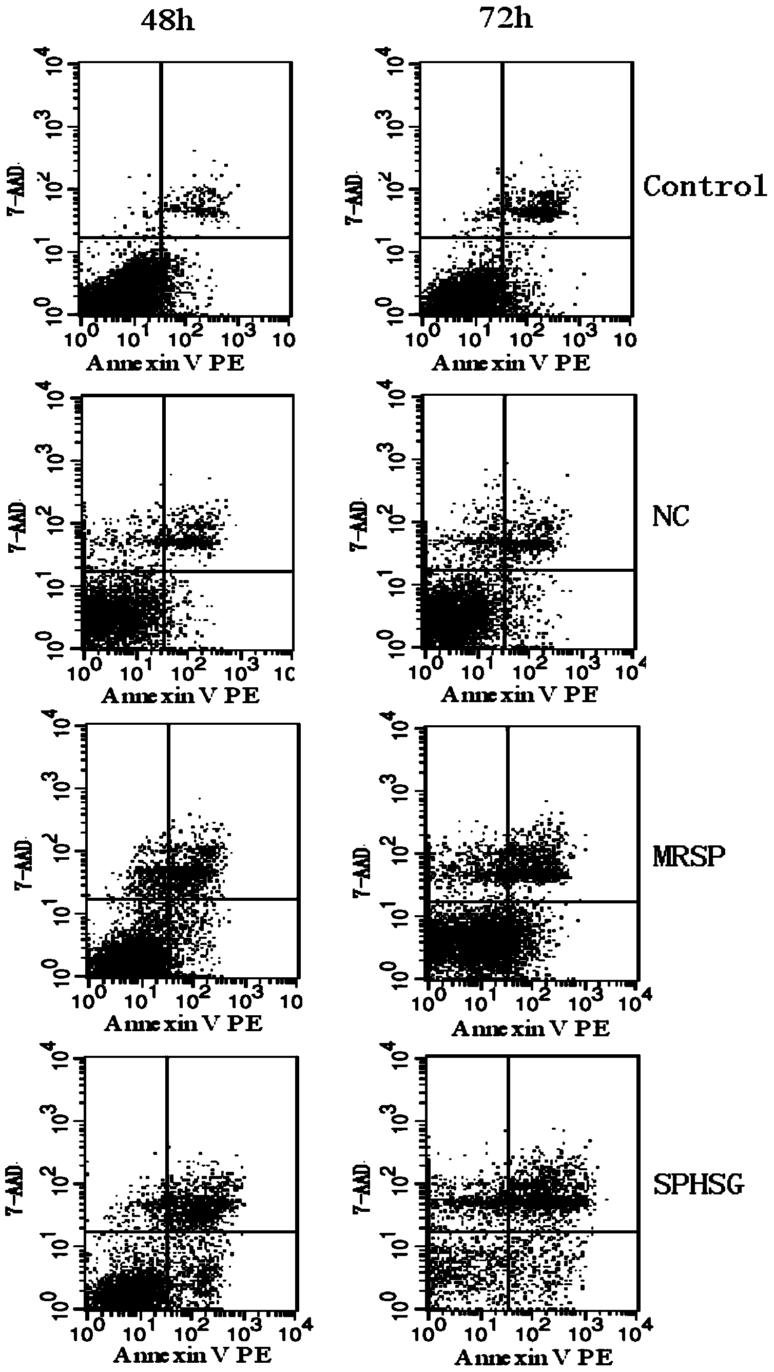
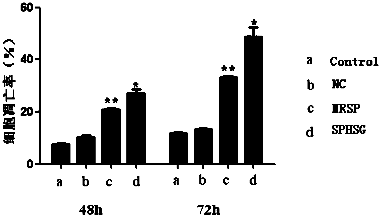
[0056] Experimental Example 1: Detection of smooth muscle cell apoptosis by flow cytometry

[0057] 1. Take the rat VSMCs of the 4th-9th generation, and count them accurately according to 5×10 5 Cells / well were seeded in a six-well plate, and 2ml of DMEM (dulbecco's modified eagle medium cell culture medium) high-glucose medium was added. Three replicate wells could be set up for each group, and the cells were incubated at 37°C and 5% CO 2 Continue culturing for 24 hours in a cell culture incubator.

[0058] 2. Synchronize, discard the medium in each experimental well, wash with sterile PBS (phosphate buffer solution) twice, pay attention to the operation should be gentle, try to avoid blowing up the cells, and then use a pipette gun to respectively Add 2ml of serum-free DMEM medium to each experimental well. Add SPHSG, MRSP and NC (invalid control) to each experimental group at a concentration of 25 μmol / L, and only add serum-free medium to the blank group, mix well and pla...

experiment example 2

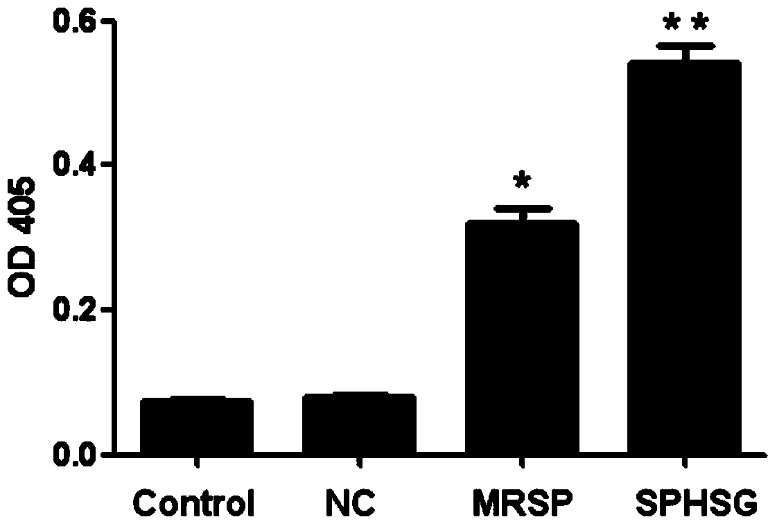
[0064] Experimental Example 2: Detection of Caspase3 Enzyme Activity

[0065] 1. Take the rat VSMCs of the 4th-9th generation, and count them accurately according to 1×10 6 Seed each cell / well in a 60mm cell culture dish, add 3ml DMEM high-glucose medium, set up 3 replicate wells for each group, and incubate at 37°C, 5% CO 2 Continue culturing for 24 hours in a cell culture incubator.

[0066] 2. Synchronize, discard the medium in each experimental well, wash with sterile PBS twice, pay attention to the operation to be gentle, try to avoid blowing up the cells, and then add 3ml serum-free DMEM to each experimental well with a pipette gun Medium. SPHSG, MRSP and NC were added to each experimental group at a concentration of 25 μmol / L, and only serum-free medium was added to the blank group, mixed evenly and placed in a cell culture incubator for 8 hours.

[0067] 3. Collect the cells when the cells have not entered the late stage of apoptosis, and collect the cells completel...

experiment example 3

[0076] Experimental example 3: In situ terminal transferase labeling technique (TUNEL) detection of apoptosis rate

[0077] 1. For cell slides, soak the autoclaved glass slides with 10% poly-slothine, air-dry them under sterile conditions, transfer them to a six-well plate, and set aside.

[0078] 2. Take the rat VSMCs of the 4th-9th generation, and count them accurately according to 2×10 5 Cells / well were inoculated in a six-well plate, 2ml DMEM high-glucose medium was added, and three replicate wells could be set up for each group, and the 2 Continue culturing for 24 hours in a cell culture incubator.

[0079] 3. Synchronize, suck up and discard the medium in each experimental well, wash with sterile PBS twice, pay attention to the operation to be gentle, try to avoid blowing up the cells, and then add 2ml serum-free DMEM to each experimental well with a pipette gun Medium. SPHSG, MRSP and NC were added to each experimental group at a concentration of 25 μmol / L, and only ...


PUM


Abstract

The invention discloses a polypeptide for inhibiting cell proliferation and inducing apoptosis and a preparation method and use thereof. The polypeptide disclosed by the invention comprises a polypeptide sequence 'SPHSG', namely YGRKKRRQRRR-GYLSKVRGISEVL, or another polypeptide sequence 'MRSP', which is similar to the polypeptide sequence in homology and function, namely YGRKKRRQRRR-DVKGYLSKVRGISEVL. An experimental basis is provided for application of the polypeptide in a coated balloon or a coated bracket. Compared with the traditional coated drug, the polypeptide does not have toxic or side effects and immunogenicity of chemical drugs as a small segment of Mfn2. The practicability is also superior to rapamycin. Therefore, the polypeptide has a wide application prospect and great clinical application value.

Description


Claims


Application Information

Patent Timeline
Owner TONGJI HOSPITAL ATTACHED TO TONGJI MEDICAL COLLEGE HUAZHONG SCI TECH
Why Eureka
Try Eureka