Automatic bovine cell factor chemiluminescence immunoassay detection method based on magnetic particles

A chemiluminescence immunoassay and magnetic particle technology, applied in the field of chemiluminescence immunoassay, can solve the problems of poor repeatability, susceptibility to interference by other cytokines, time-consuming and other problems, achieve large surface area, speed up analysis and detection time, easy to capture and clean Effect

Inactive Publication Date: 2014-03-19
YANGZHOU UNIV
View PDF4 Cites 8 Cited by
  • Summary
  • Abstract
  • Description
  • Claims
  • Application Information

AI Technical Summary

Problems solved by technology

Traditional methods for detecting bovine IFN-γ and IL-4 are mostly biological methods, but they are time-consuming, poor in repeatability, and susceptible to interference from other cytoki

Method used


Image

  • Automatic bovine cell factor chemiluminescence immunoassay detection method based on magnetic particles
  • Automatic bovine cell factor chemiluminescence immunoassay detection method based on magnetic particles
  • Automatic bovine cell factor chemiluminescence immunoassay detection method based on magnetic particles

Examples


Embodiment 1

[0031] (1) Preparation of immunosensor

[0032] Take the magnetic bead stock solution, wash the magnetic beads with the activation buffer solution, and then disperse them in the activation buffer solution, add NHS and EDC solids to it in sequence, and keep stirring at room temperature for activation. After the activation is completed, wash with PBS buffer solution and disperse in the bovine IFN-γ antibody solution, stir at room temperature for a certain period of time, and then fix at 4°C overnight. After the fixation is completed, the magnetic beads are washed with PBS buffer solution, and then blocked by adding a blocking solution, and the preparation of the immunosensor is completed.

[0033] (2) Analysis steps

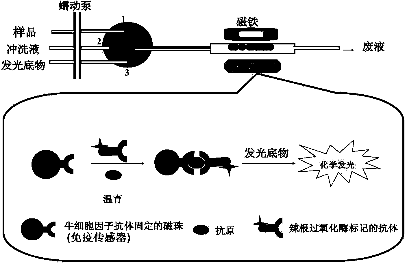
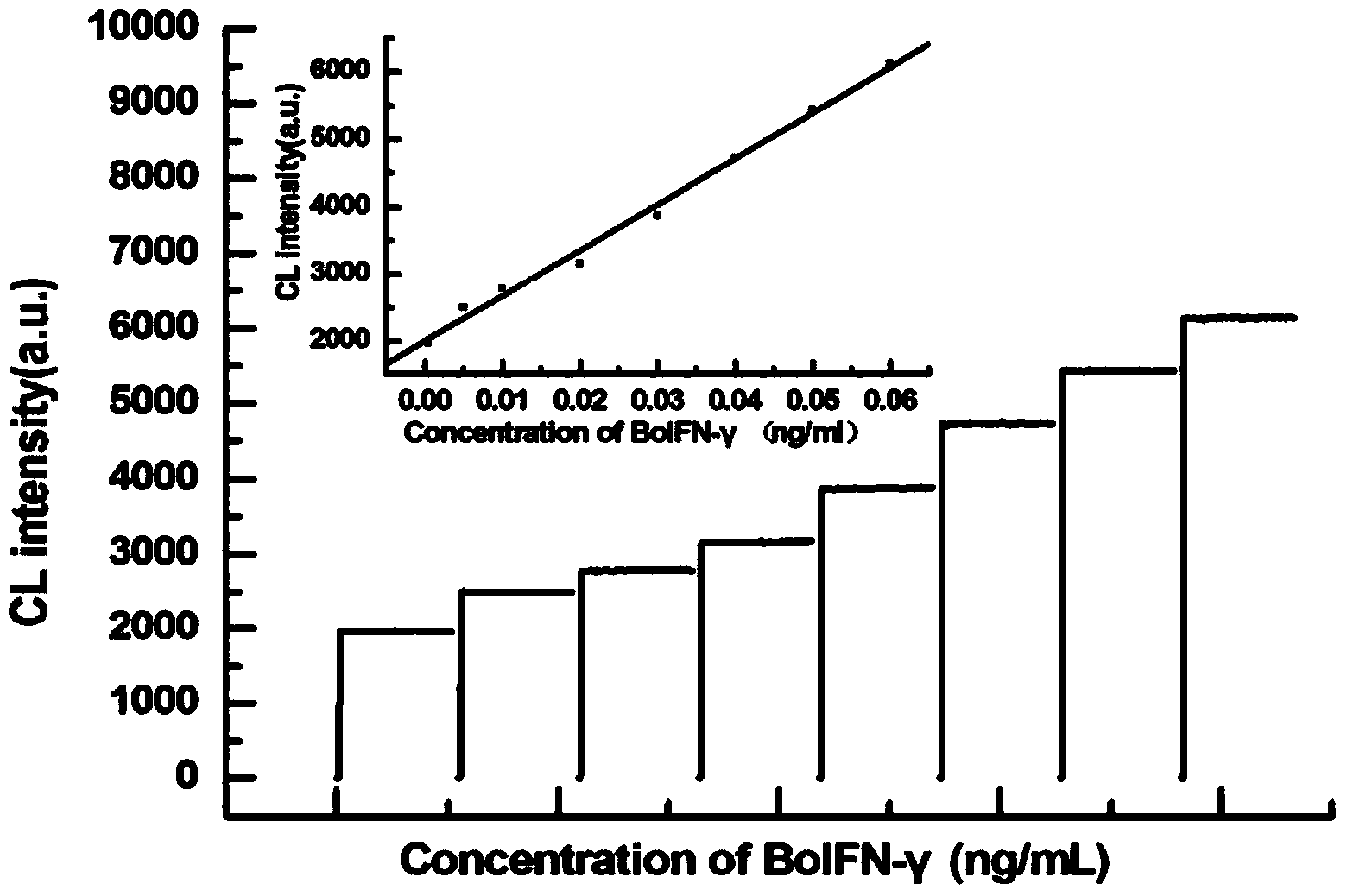
[0034] Analysis diagram as figure 1 , the magnetic bead dispersion immobilized with bovine IFN-γ antibody, bovine IFN-γ antigen solution, and horseradish peroxidase-labeled antibody were mixed and injected into a glass tube, and the three formed a sandwich comple...

Embodiment 2

[0035] (1) Preparation of immunosensor

[0036] Take the magnetic bead stock solution, wash the magnetic beads with the activation buffer solution, and then disperse them in the activation buffer solution, add NHS and EDC solids to it in sequence, and keep stirring at room temperature for activation. After the activation is completed, wash with PBS buffer solution and disperse in the bovine IL-4 antibody solution, stir at room temperature for a certain period of time, and then fix at 4°C overnight. After the fixation is completed, the magnetic beads are washed with PBS buffer solution, and then blocked by adding a blocking solution, and the preparation of the immunosensor is completed.

[0037] (2) Analysis steps

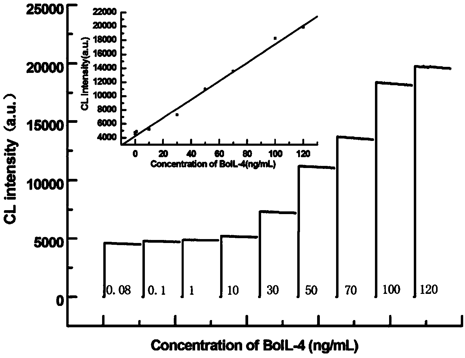
[0038] The magnetic bead dispersion immobilized with bovine IL-4 antibody, bovine IL-4 antigen solution, and horseradish peroxidase-labeled antibody were mixed and injected into a glass tube, and the three formed a sandwich complex during incubation. Sandwich comp...


PUM

PropertyMeasurementUnit
The inside diameter ofaaaaaaaaaa
Particle sizeaaaaaaaaaa

Abstract

The invention relates to an automatic bovine cell factor chemiluminescence immunoassay detection method based on magnetic particles. Due to a sandwich immunoassay method, a magnetic ball serves as a fixed carrier of an antibody, and flow injection is combined to be applied to chemiluminescence analysis, so that the high-sensitivity automatic bovine cell factor chemiluminescence immunoassay detection method is built. An immunosensor is manufactured by activating the magnetic ball with the carboxyl, then covalently connecting the antibody to the surface of the magnetic ball and closing residual active sites; when being applied to detection of bovine cell factors, the immunosensor is fed into an immune micro reactor after being mixed with an antigen and the antibody which is labeled with horseradish peroxidase; the immunosensor is incubated on line to form a sandwich compound, and the sandwich compound is fed into a chemiluminiscence substrate; a luminous signal is immediately acquired, so that quick detection can be realized. The automatic bovine cell factor chemiluminescence immunoassay detection method has the characteristics of short analysis time, simplicity in operation, high specificity, high sensitivity and the like and can be used for quantitative detection of the bovine cell factors and researches on immunomechanisms of the bovine cell factors.

Description

technical field [0001] The invention relates to an automatic bovine cytokine chemiluminescence immunodetection method based on magnetic microparticles, which is characterized in that magnetic microbeads are used as antibody immobilization carriers to prepare bovine cytokine immunosensors, which are combined with flow injection to construct a highly sensitive An automatic chemiluminescence immunoassay method for quickly detecting bovine cytokines belongs to the field of chemiluminescence immunoassay. Background technique [0002] Cytokines are small molecular soluble proteins that are synthesized and secreted by various cells in the body and have various physiological activities and participate in pathological reactions. It plays a fundamental supervisory and signaling role in the immune response. Divided into interferon, interleukin, tumor necrosis factor, chemokine, colony-stimulating factor, growth factor, etc. Bovine cytokines mainly include bovine gamma interferon (IFN...

Claims


Application Information

Patent Timeline
IPC IPC(8): G01N33/68
CPCG01N33/68G01N33/54326
Inventor 杨占军罗淑芬张琼陈祥
Owner YANGZHOU UNIV
Why Eureka
Try Eureka