Method and product for localised or spatial detection of nucleic acid in a tissue sample

A tissue sample, local detection technology, which is applied in the preparation of test samples, biochemical equipment and methods, and the determination/inspection of microorganisms, etc., can solve the problem of no research on high-throughput methods

Active Publication Date: 2014-05-07
十程基因技术瑞典公司
View PDF9 Cites 17 Cited by
  • Summary
  • Abstract
  • Description
  • Claims
  • Application Information

AI Technical Summary

Problems solved by technology

However, to date, there is no high-throughput method for studying transcriptional activity in intact tissues at high resolution

Method used


Image

  • Method and product for localised or spatial detection of nucleic acid in a tissue sample
  • Method and product for localised or spatial detection of nucleic acid in a tissue sample
  • Method and product for localised or spatial detection of nucleic acid in a tissue sample

Examples


Embodiment 1

[0359] Array preparation

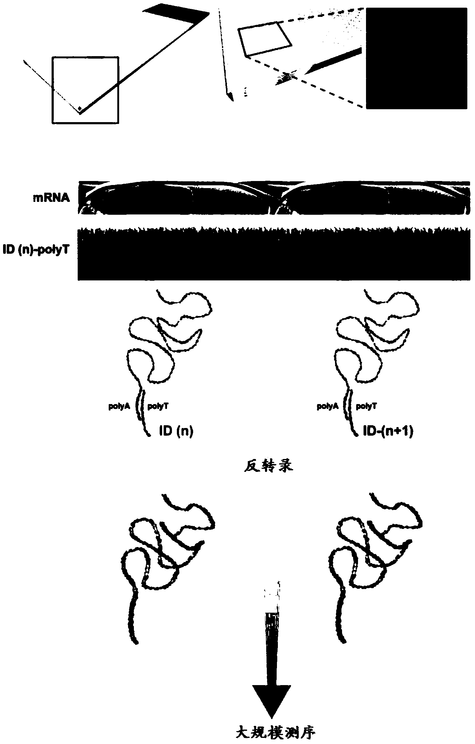
[0360] The following experiments demonstrate how to attach oligonucleotide probes to array substrates via their 5' or 3' ends to obtain arrays containing capture probes that hybridize to mRNA.

[0361] Preparation of self-made spray-dot microarray with probes in 5' to 3' direction

[0362] Twenty RNA capture oligonucleotides (labeled 1-20, Table 1) each with a labeled sequence were spotted on a glass slide as capture probes. The probe was synthesized with a 5'-terminal amino linker and a C6 spacer. All probes were synthesized by Sigma-Aldrich (St. Louis, MO, USA). The RNA capture probe was prepared with 150 mM sodium phosphate to make a suspension with a concentration of 20 μM and a pH of 8.5, and the Nanoplotter NP2.1 / E spraying system (Gesim, Grosserkmannsdorf, Germany) was used in CodeLink TM Activated microarray slides (7.5cm x 2.5cm; Surmodics, Eden Prairie, MN, USA) were spotted. After spraying is complete, follow the manufacturer's inst...

Embodiment 2

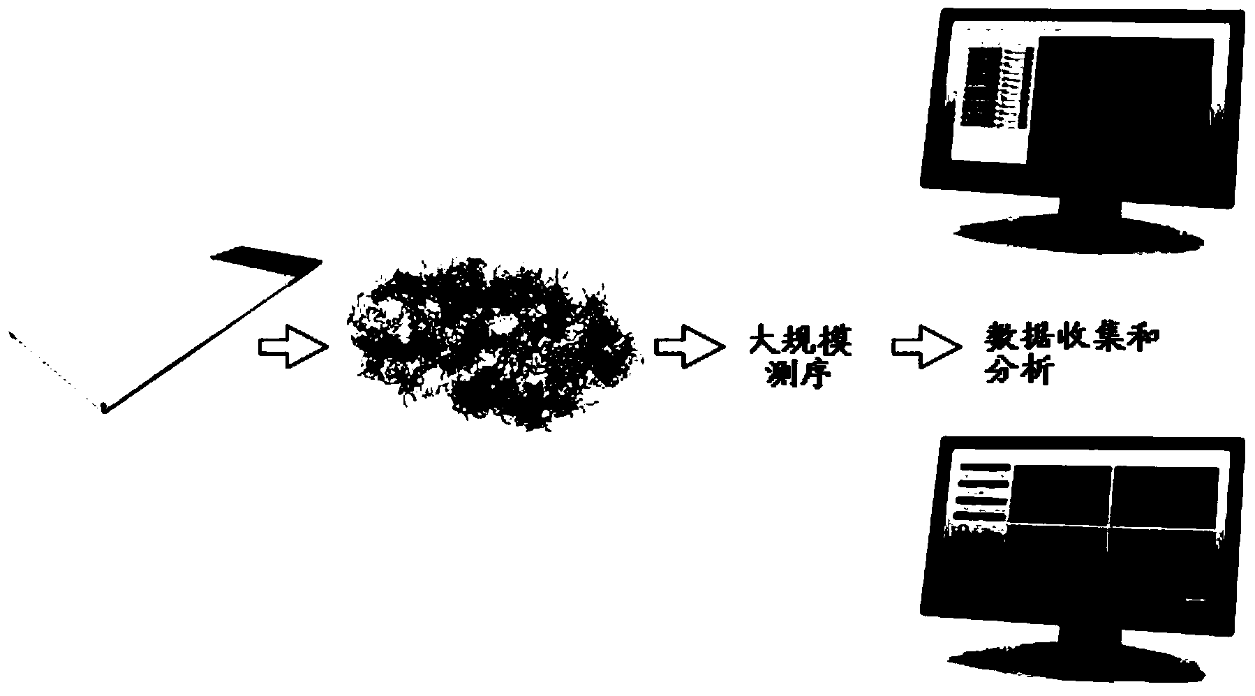
[0433] Figure 8 to Figure 12 Demonstrates successful visualization of stained FFPE mouse brain tissue (olfactory bulb) sections placed on transcriptome capture arrays labeled with identifiers, following the general protocol described in the Examples. Compared with the experiments of fresh frozen tissue in Example 1, Figure 8 The FFPE tissue morphology shown is better. Figure 9 and Figure 10 Shows how to localize tissue on arrays of different probe density types.

Embodiment 3

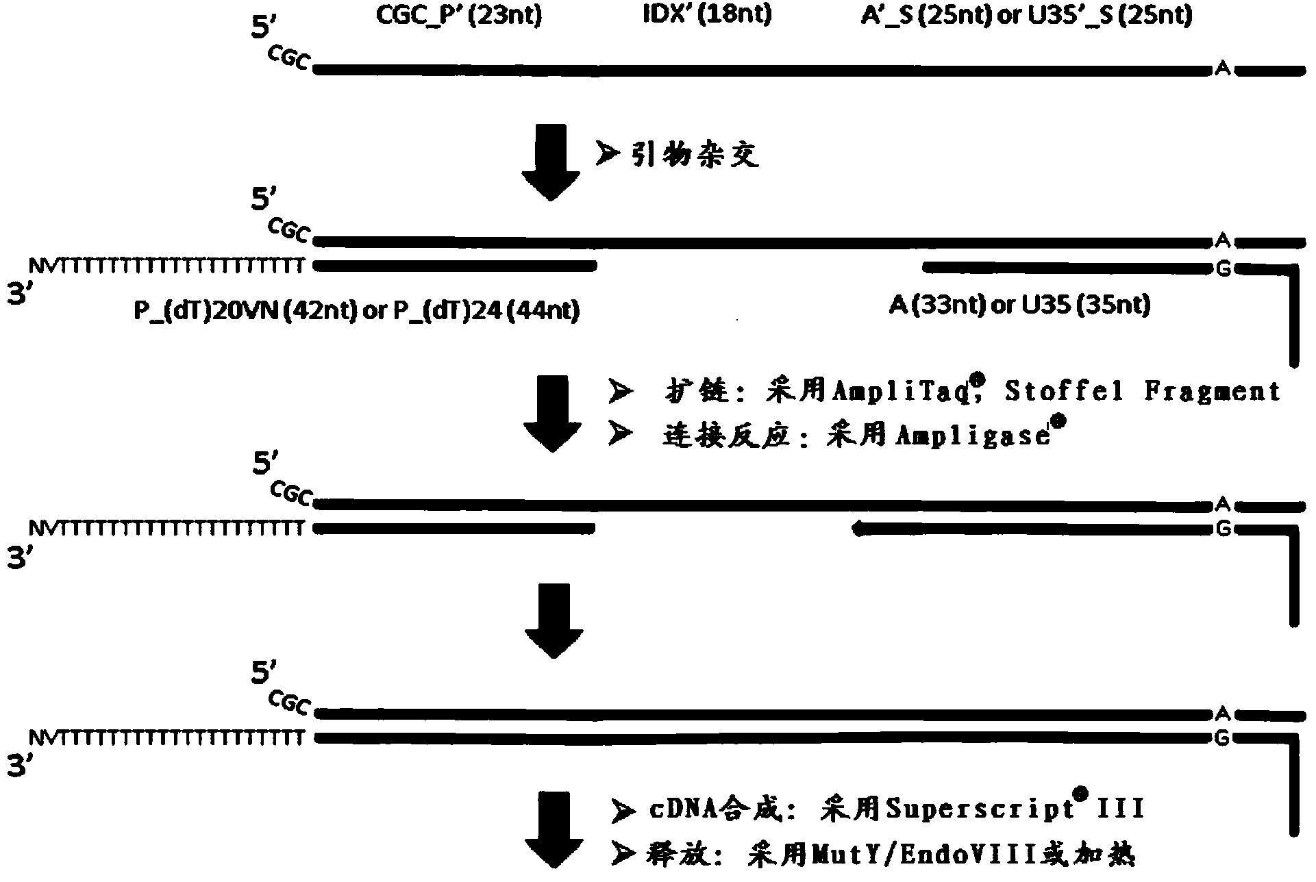
[0435] Whole-transcriptome amplification with random-primed second-strand synthesis and universal-end amplification (capture probes containing tag sequences retain at the end of the resulting dsDNA)

[0436] After release of capture probes with PCR buffer containing USER enzyme mix capable of cleaving uracil (covalently attached probes)

[0437] or

[0438] After release of capture probes with heated PCR buffer (synthetic capture probes for in situ hybridization)

[0439] Add 40μl of 1x containing 1.8mM MgCl to each of the 2 tubes 2 Fast start high-fidelity PCR buffer (Faststart HiFi PCR Buffer with1.8mM MgCl 2 ) (pH8.3) (Roche, www.roche-applied-science.com), dNTP 0.2mM each (Fermentas, www.fermentas.com), 0.1μg / μl BSA (New England Biolabs, www.neb.com) , 0.1 U / μl USER enzyme (New England Biolabs), released cDNA (extended from surface probe) and released surface probe, and 1 μl RNaseH (5 U / μl). Then, 2 tubes were incubated in a thermal cycler (Applied Biosystems, www....


PUM


Abstract

The present invention relates to methods and products for the localised or spatial detection of nucleic acid in a tissue sample and in particular to a method for localised detection of nucleic acid in a tissue sample comprising: (a) providing an array comprising a substrate on which multiple species of capture probes are directly or indirectly immobilized such that each species occupies a distinct position on the array and is oriented to have a free 3' end to enable said probe to function as a primer for a primer extension or ligation reaction, wherein each species of said capture probe comprises a nucleic acid molecule with 5' to 3': (i) a positional domain that corresponds to the position of the capture probe on the array, and (ii) a capture domain; (b) contacting said array with a tissue sample such that the position of a capture probe on the array may be correlated with a position in the tissue sample and allowing nucleic acid of the tissue sample to hybridise to the capture domain in said capture probes; (c) generating DNA molecules from the captured nucleic acid molecules using said capture probes as extension or ligation primers, wherein said extended or ligated DNA molecules are tagged by virtue of the positional domain; (d) optionally generating a complementary strand of said tagged DNA and / or optionally amplifying said tagged DNA; (e) releasing at least part of the tagged DNA molecules and / or their complements or amplicons from the surface of the array, wherein said part includes the positional domain or a complement thereof; and (f) directly or indirectly analysing the sequence of the released DNA molecules.

Description

technical field [0001] The present invention is primarily concerned with the localized or spatial detection of nucleic acids in tissue samples. The nucleic acid may be RNA or DNA. Therefore, the methods provided by the present invention can be used to detect and / or analyze RNA (such as RNA transcripts) or genomic DNA to obtain spatial information about gene localization, distribution or expression in tissue samples (such as individual cells), or specifically , the spatial information on the location or distribution of any genomic variation (not necessarily a variation in a gene). Therefore, the present invention enables the study of spatial genomics and spatial transcriptomics. [0002] A quantitative and / or qualitative method capable of analyzing the distribution, location or expression of genomic sequences in a tissue sample in which spatial expression, distribution or localization patterns are preserved. Thus, the new method provides a pipeline for conducting "spatial tr...

Claims


Application Information

Patent Timeline
Patent Type & Authority Applications(China)
IPC IPC(8): C12Q1/68
CPCC12Q1/6837C12Q1/6841C12Q1/6844C12Q2543/101C12Q2565/514C12Q2565/537C12Q1/6853C12Q2563/179C12N15/11C12Q1/6806G16B50/30G16B50/20G16B30/00C12Q1/6816C12Q1/6827C12Q1/6876C12Y600/00C12N15/1065C12Q1/682G01N1/30G01N1/42
Inventor 乔纳斯·弗瑞森帕特里克·斯塔尔乔亚基姆·伦德贝格
Owner 十程基因技术瑞典公司
Why Eureka
Try Eureka