Method for segmenting inhomogeneous medical image
A medical image, uniform technology, applied in the field of image analysis, can solve problems such as inability to achieve segmentation and inability to achieve segmentation
- Summary
- Abstract
- Description
- Claims
- Application Information
AI Technical Summary
Problems solved by technology
Method used
Image
Examples
example 1
[0039] Example 1 (Segmentation of non-uniform 2D medical images)
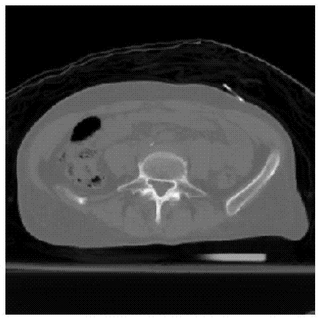
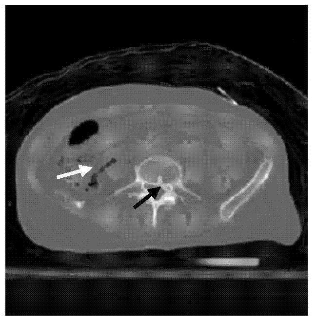
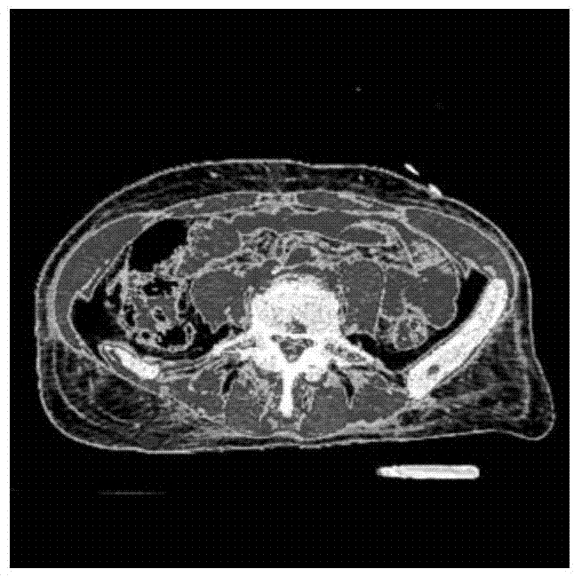
[0040] This embodiment takes figure 1 The illustrated abdominal CT (Computed Tomography) image of a certain patient is taken as an example to describe the implementation process of the method of the present invention. figure 1 The size of is 512·512, and the lumbar vertebrae to be segmented belong to heterogeneous targets, which include high-density cortical bone, medium-density cortical bone and low-density bone marrow. The specific segmentation method is as follows:
[0041] Step 1: Read in as figure 1 For the CT image shown, through the MATLAB GUI graphical interface, select the foreground seed points that can represent the lumbar vertebrae and the background seed points that represent other normal abdominal tissues. Overlay the selected seed points on figure 1 on, get as figure 2 For the abdominal CT image of the lumbar vertebrae to be segmented in the marked seed point set shown, the gray information...
example 2
[0050] Example 2 (Segmentation of non-uniform 3D medical images)
[0051] This embodiment takes Figure 8 The pelvic CT image containing the applicator taken by a certain cervical cancer patient receiving brachytherapy is taken as an example to describe the method of the present invention for the non-uniform three-dimensional medical image segmentation process. Figure 8 The size of the source is 256·256·55, and the source applicator in the figure is a non-uniform three-dimensional object, which includes high-density metal tubes, medium-density plastics, and low-density liquids and air. The specific segmentation method is as follows:
[0052] Step 1: Read in as Figure 8 For the pelvic CT image shown, select the foreground seed point representing the applicator and the background seed point representing other pelvic tissue structures through the MATLAB GUI graphical interface. Overlay the selected seed points on Figure 8 on, get as Figure 9 The pelvic CT image containin...
PUM
 Abstract
Description
Claims
Application Information
 - R&D
- Intellectual Property
- Life Sciences
- Materials
- Tech Scout
- Unparalleled Data Quality
- Higher Quality Content
- 60% Fewer Hallucinations
Browse by: Latest US Patents, China's latest patents, Technical Efficacy Thesaurus, Application Domain, Technology Topic, Popular Technical Reports.
© 2025 PatSnap. All rights reserved.Legal|Privacy policy|Modern Slavery Act Transparency Statement|Sitemap|About US| Contact US: help@patsnap.com


