Glandular tissue characteristic gray scale detection method and device

A technology of characteristic grayscale and breast tissue, applied in image data processing, medical science, instruments, etc., can solve the problems of unrecognizable, increased exposure dose, etc., to avoid inaccurate and reasonable characteristic grayscale, and avoid secondary exposure dose Effect

Active Publication Date: 2014-06-25
NEUSOFT MEDICAL SYST CO LTD
View PDF2 Cites 20 Cited by
  • Summary
  • Abstract
  • Description
  • Claims
  • Application Information

AI Technical Summary

Problems solved by technology

This method cannot identify interference factors such as larger lesions in the gland or parapets (the gray value of these interference factors is also small), resulting in an increase in exposure dose

Method used


Image

  • Glandular tissue characteristic gray scale detection method and device
  • Glandular tissue characteristic gray scale detection method and device
  • Glandular tissue characteristic gray scale detection method and device

Examples


Embodiment 2

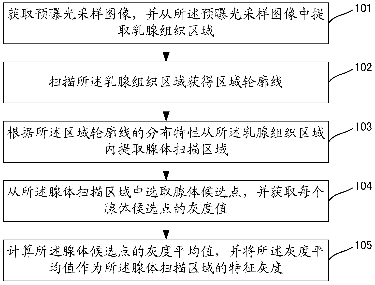
[0182] In addition, if the effective gray scale range of the breast tissue area is obtained before the breast tissue area is extracted from the pre-exposure sampling image, the present invention also provides Figure 15 The shown breast tissue area extraction unit embodiment 2 includes:

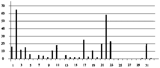
[0183] A setting unit 701, configured to set channel values ​​of channels in the grayscale histogram outside the effective grayscale range to zero after the grayscale histogram of the pre-exposure sampling image is generated;

[0184] a peak extraction unit 702, configured to extract the peak of the grayscale histogram;

[0185] A judging unit 703, configured to judge whether the number of extracted peaks is less than two;

[0186] A first determination unit 704, configured to determine the pre-exposure sampling image as the breast tissue region when the determination unit determines that the number of peaks is less than two;

[0187] The selection unit 705 is used to select the two peaks w...


PUM


Abstract

The invention discloses a glandular tissue characteristic gray scale detection method and device. The method comprises that: a pre-exposure sampling image is acquired, and a mammary tissue area is extracted from the pre-exposure sampling image; the mammary tissue area is scanned so that area contour lines are acquired; gland scanning areas are extracted from the mammary tissue area according to distribution characteristics of the area contour lines; gland candidate points are selected from the gland scanning areas, and a gray scale value of each gland candidate point is acquired; and a gray scale mean value of the gland candidate points is calculated, and the gray scale mean value acts as the characteristic gray scale of the gland scanning areas. The process has no participation of personnel so that time and labor are saved, and defects of inaccurate and unreasonable characteristic gray scale caused by artificial factors are also avoided. Besides, the defects in the prior art that secondary exposure dose is increased under the influence of interference factors can be reduced and even avoided by the technical scheme.

Description

technical field [0001] The invention relates to a grayscale detection method and device for glandular tissue features. Background technique [0002] Mammography machine is a commonly used breast examination equipment. In order to obtain breast projection images with relatively stable quality, most mammography machines use pre-exposure mode for automatic exposure control. The process can be briefly described as follows: First, through a very small dose Exposure to obtain a pre-exposure sampling image; then calculate the dose required to obtain a complete exposure according to the gray value of the pre-defined area and the target gray value; finally perform a second exposure according to the dose required for a complete exposure to obtain the mammary gland Expose the image. It can be seen from the control process that the gray value of the pre-defined area directly affects the dose required for the final complete exposure, and then affects the image quality of the second expo...

Claims


Application Information

Patent Timeline
Patent Type & Authority Applications(China)
IPC IPC(8): G06T7/00A61B6/00
Inventor 李海春
Owner NEUSOFT MEDICAL SYST CO LTD
Why Eureka
Try Eureka