Ultrasound breaking method for animal tissue in chromatin co-immunoprecipitation
A technology of co-immunoprecipitation and animal tissue, which is applied in the direction of biochemical equipment and methods, and the determination/inspection of microorganisms, to achieve the effects of saving consumption, saving consumables, and reducing time consumption
- Summary
- Abstract
- Description
- Claims
- Application Information
AI Technical Summary
Benefits of technology
Problems solved by technology
Method used
Image

Examples
Embodiment 1
[0034] Example 1 Ultrasonic disruption method of animal tissue in chromatin immunoprecipitation (taking chicken embryo as an example)
[0035] 1. ChIP experimental sample preparation
[0036] After the collected eggs were sterilized, they were placed in an incubator with a temperature of 37.8°C and a humidity of 60%, and the eggs were turned every 2 hours. After incubation until the 16th day, the heart tissue of the chicken embryos was collected and quickly frozen in liquid nitrogen.
[0037] 2. Exploration of ultrasonic crushing experimental conditions and result verification in the ChIP experimental process
[0038] The reagents used in the experiments were from ChIP unless otherwise noted. TM G Tissue Kit (Millipore, USA)
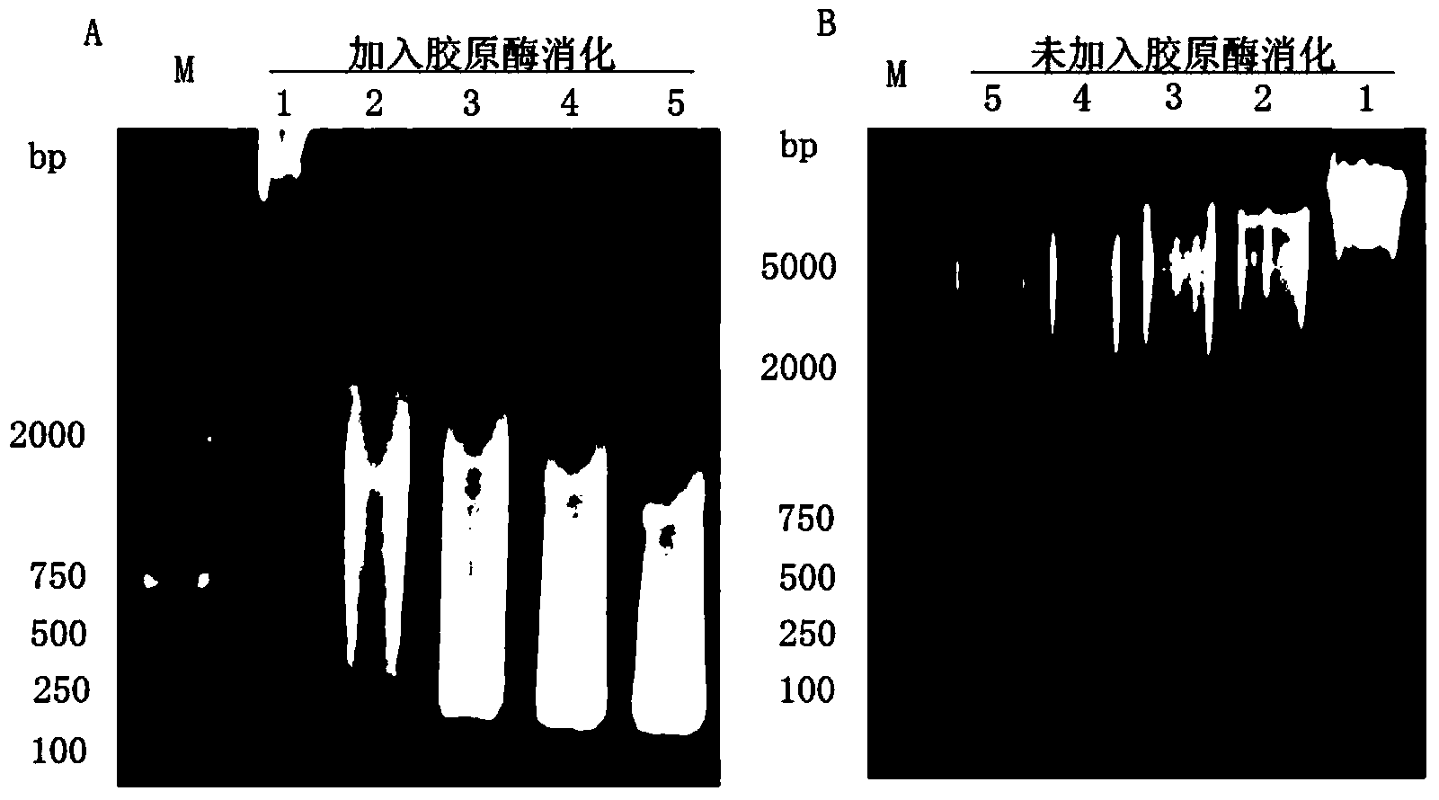
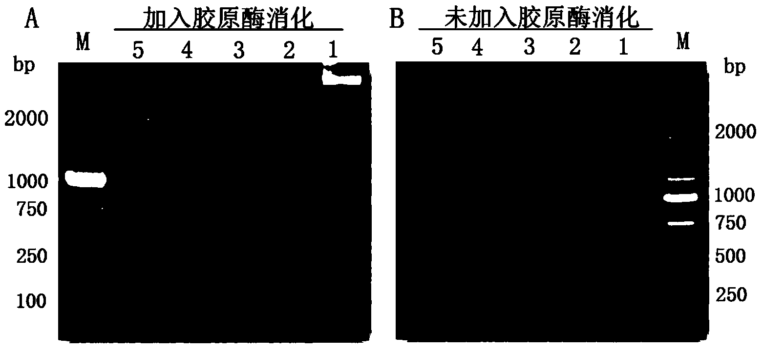
[0039] Take about 50ug of frozen chicken embryo heart tissue, thaw slowly on ice and chop into pieces → add 1ml 1:1 mixed collagenase (Sigma, C0130, USA), DPBS (Gibco, C14190, USA) solution, and digest the animal in a shaking water bath at 37°C Centr...
Embodiment 2
[0043] Example 2 Ultrasonic disruption method of animal tissue in chromatin immunoprecipitation (taking pig tissue as an example)
[0044] 1. ChIP experimental sample preparation
[0045] Heart tissue from 6-month-old pigs was collected and quickly frozen in liquid nitrogen.
[0046] 2. Exploration of ultrasonic crushing experimental conditions and result verification in the ChIP experimental process
[0047] The reagents used in the experiments were from ChIP unless otherwise noted. TM G Tissue Kit (Millipore, USA)
[0048] Take about 50ug of frozen pig heart tissue, thaw slowly on ice and chop into pieces → add 1ml of 1:1 mixed collagenase (Sigma, C0130, USA), DPBS (Gibco, C14190, USA) solution, and digest the animal tissue in a shaking water bath at 37°C 3.5h→4℃, centrifuge at 800g for 15min, remove supernatant→add 500ul TSS / PI, pipette repeatedly with pipette tip→4℃, centrifuge at 800g for 5min, remove supernatant→add 1ml1×phosphate buffer saline (PBS) / 1% Formaldehyde...
PUM
 Abstract
Description
Claims
Application Information
 - R&D
- Intellectual Property
- Life Sciences
- Materials
- Tech Scout
- Unparalleled Data Quality
- Higher Quality Content
- 60% Fewer Hallucinations
Browse by: Latest US Patents, China's latest patents, Technical Efficacy Thesaurus, Application Domain, Technology Topic, Popular Technical Reports.
© 2025 PatSnap. All rights reserved.Legal|Privacy policy|Modern Slavery Act Transparency Statement|Sitemap|About US| Contact US: help@patsnap.com