A method and device for locating grayscale image regions when displaying medical images

A gray-scale image and medical image technology, applied in the field of medical displays, can solve problems such as misjudgment, image error correction, and excessive brightness, and achieve the effects of improving precision and accuracy, accurate image range, and improving accuracy

Active Publication Date: 2017-02-15
HISENSE VISUAL TECH CO LTD
View PDF5 Cites 0 Cited by
  • Summary
  • Abstract
  • Description
  • Claims
  • Application Information

AI Technical Summary

Problems solved by technology

However, since the values ​​of the three RGB components of pixels displaying black and white text or black and white images are equal, the black and white text (such as signs) in the color image or the area of ​​the black and white image will be wrongly determined as gray scale by the above method area, so that DICOM correction is performed on the black and white text or black and white image in the color image, so that the black and white text or image in the color image will appear too bright, distorted, etc., and it may also cause doctors or users to display the so-called gray scale. Misjudgment of image content
It can be seen that because the black and white text or the black and white image area in the color image is mislocated as a gray scale area, the correction of the image error is caused, so that the high quality display of the gray scale image and the color image at the same time cannot be satisfied by a single display.

Method used


Image

  • A method and device for locating grayscale image regions when displaying medical images
  • A method and device for locating grayscale image regions when displaying medical images
  • A method and device for locating grayscale image regions when displaying medical images

Examples


Embodiment Construction

[0023] The following will clearly and completely describe the technical solutions in the embodiments of the present invention with reference to the accompanying drawings in the embodiments of the present invention. Obviously, the described embodiments are only some, not all, embodiments of the present invention. Based on the embodiments of the present invention, all other embodiments obtained by persons of ordinary skill in the art without creative efforts fall within the protection scope of the present invention.

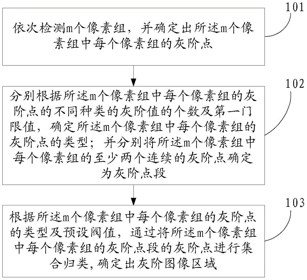
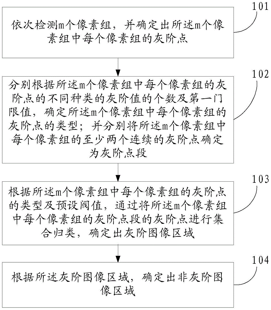
[0024] An embodiment of the present invention provides a method for locating grayscale image regions when displaying medical images, such as figure 1 shown, including:

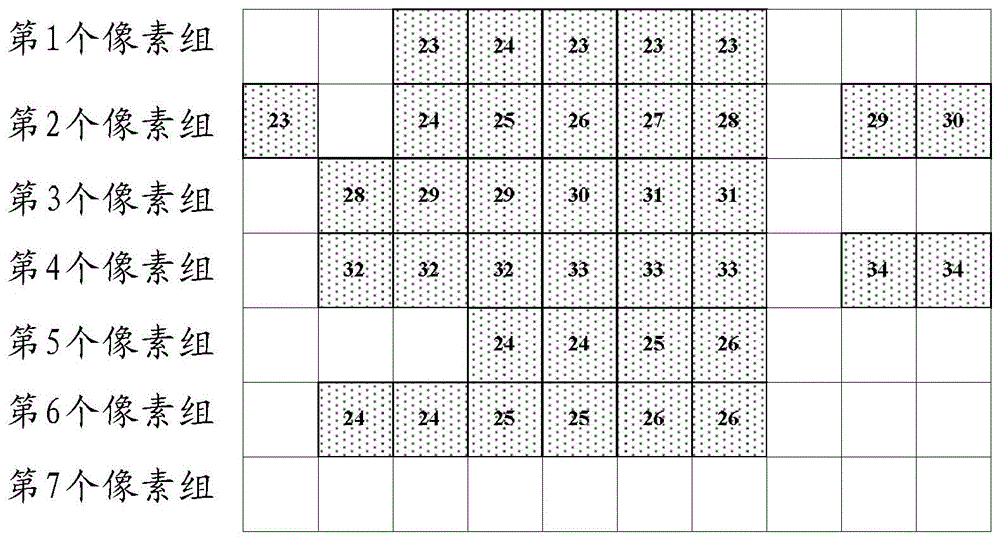
[0025] 101. Detect m pixel groups in sequence, and determine the grayscale point of each pixel group in the m pixel groups.

[0026] Wherein, the m is an integer greater than 0. Each pixel group includes at least one pixel.

[0027] Specifically, after the device for locating the grayscale image...


PUM


Abstract

An embodiment of the invention provides a method and apparatus for positioning a gray scale image region during medical image displaying and relates to the field of medical displays. The method and apparatus for positioning the gray scale image region during medical image displaying is used for improving the accuracy of image correction through the improvement of the accuracy of positioning of the image gray scale region so as to achieve high-quality display of a gray scale image and a color image through a single display. The method includes the steps of sequentially detecting m pixel groups and determining gray scale points of every pixel group of the m pixel groups; determining types of the gray scale points of every pixel group of the m pixel groups respectively according to the number of values of the types of the gray scale points of every pixel group of the m pixel groups and a first threshold value; respectively determining at least two consecutive gray scale points of every pixel group of the m pixel groups as a gray scale point segment; gathering and classifying gray scale points of the gray scale point segment of every pixel group of the m pixel groups for determining the gray scale image region according to the types of the gray scale points of every pixel group of the m pixel groups and a preset threshold.

Description

technical field [0001] The invention relates to the field of medical displays, in particular to a method and device for locating grayscale image regions when displaying medical images. Background technique [0002] The monitor satisfies the needs of people for picture display in their life and work. Depending on the scene used by the display, the requirements for image display are different, and the correction methods used for the displayed image are different. For example: diagnostic monitors are used to display grayscale images, which often require high resolution, high contrast, high brightness, and DICOM (Digital Imaging and Communications in Medicine, medical digital imaging and communication) correction; operating room monitors face the endoscope And other front-end imaging equipment, used to display color images such as surgical auxiliary information, requires moderate resolution, general brightness, and GAMMA (gamma) correction. [0003] In some scenarios, it is ne...

Claims


Application Information

Patent Timeline
Patent Type & Authority Patents(China)
IPC IPC(8): G09G5/00G09G5/02
Inventor 骆俊谕高维嵩孟现策
Owner HISENSE VISUAL TECH CO LTD
Why Eureka
Try Eureka