Supercharge Your Innovation With Domain-Expert AI Agents!

A method for extracting and marking medical image feature area in real time

A feature area and medical image technology, applied in the field of medical image analysis, can solve the problems of no feature area prompt, inconvenient observation, easy to ignore feature area, etc., to achieve the effect of improving efficiency and saving search time

Inactive Publication Date: 2015-02-04
SHANGHAI QIZHENG MICROELECTRONICS
View PDF5 Cites 11 Cited by
  • Summary
  • Abstract
  • Description
  • Claims
  • Application Information

AI Technical Summary

Problems solved by technology

[0006] In the above method, the doctor does not have a reminder of the characteristic area during the collection and observation process, it is easy to ignore the characteristic area, and it is not easy to observe

Method used


Image

  • A method for extracting and marking medical image feature area in real time
  • A method for extracting and marking medical image feature area in real time
  • A method for extracting and marking medical image feature area in real time

Examples


Embodiment Construction

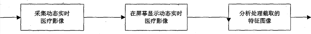
[0038] The method for real-time extraction and labeling of medical image feature regions of the present invention comprises the following steps:

[0039] (1) Acquisition of dynamic real-time medical images;

[0040] (2) Display dynamic real-time medical images on the screen, and mark the feature area (the feature area can be set as a lesion area, suspected lesion area or observed object area according to needs).

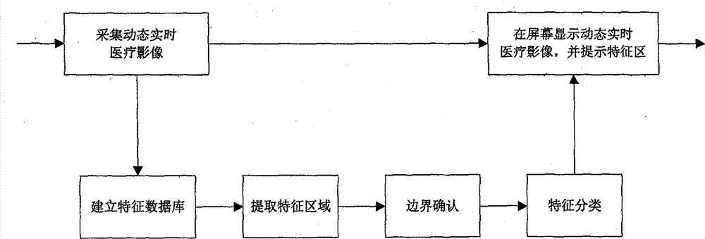
[0041] Preferably, as figure 2 As shown, in the method for real-time extraction and labeling of medical image feature regions of the present invention, the synchronization between the step (1) and the step (2) includes the following steps:

[0042] (21) Establish a feature database;

[0043] (22) extract feature region;

[0044] (23) Boundary confirmation;

[0045] (24) Feature classification.

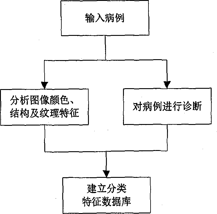
[0046] Such as image 3 As shown, the medical image feature region real-time extraction and labeling method of the present invention, the step (21) establishes a featu...


PUM


Abstract

The invention discloses a method for extracting and marking a medical image feature area in real time, the method comprising steps of: 1) collecting a dynamic real-time medical image; 2)displaying on a screen the dynamic real time medical image, and marking a feature area. The method for extracting and marking the medical image feature area in real time of the invention can implement automatic capturing of a feature area (a focal area, a focal suspected area, a key observation area) in real time, marking a corresponding local area to assist human eyes of a doctor in distinguishing, and thus the invention saves searching time and improves greatly the efficiency of surgery.

Description

technical field [0001] The invention relates to a medical image analysis method used in surgical medicine for internal inspection, diagnosis and treatment of a human body, and specifically designs a method for real-time extraction and labeling of medical image feature regions. Background technique [0002] Equipment such as endoscopes, ultrasound machines, and CT are common medical equipment, and the medical images output by them can facilitate doctors to observe images of non-visible areas of patients (such as stomach, small intestine, abdomen, brain, lungs, kidneys and other organs), However, the analysis and processing process of existing medical images is generally operated on the image at the back end, not real-time, such as figure 1 shown, including the following steps: [0003] (1) Acquisition of dynamic real-time medical images; [0004] (2) Display dynamic real-time medical images on the screen; [0005] (3) Analyze and process the intercepted feature image. [...

Claims


Application Information

Patent Timeline
Patent Type & Authority Applications(China)
IPC IPC(8): G06K9/46G06F17/30
CPCG06F16/583G06V10/462
Inventor 钱大宏
Owner SHANGHAI QIZHENG MICROELECTRONICS
Features
  • R&D
  • Intellectual Property
  • Life Sciences
  • Materials
  • Tech Scout
  • Unparalleled Data Quality
  • Higher Quality Content
  • 60% Fewer Hallucinations
Learn More