Lung 4D-CT image exhaling process middle phase image reconstruction method based on registration

A 4D-CT and phase image technology, applied in the field of medical image processing, can solve problems such as the impact of CT image quality, achieve important application value, and reduce the effect of imaging radiation dose

Inactive Publication Date: 2015-02-18
SOUTHERN MEDICAL UNIVERSITY
View PDF0 Cites 12 Cited by
  • Summary
  • Abstract
  • Description
  • Claims
  • Application Information

AI Technical Summary

Problems solved by technology

In addition, whether the real-time phase processing system (RPM) is accurately synchronized with

Method used


Image

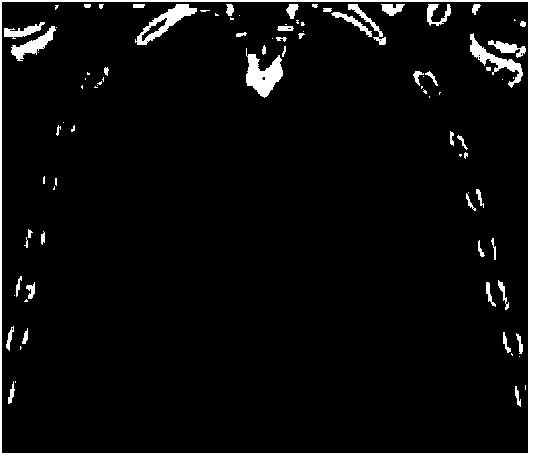
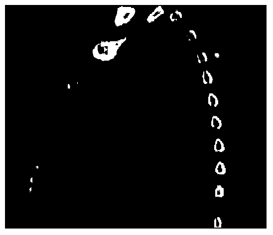
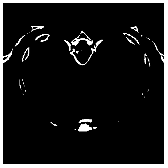
  • Lung 4D-CT image exhaling process middle phase image reconstruction method based on registration
  • Lung 4D-CT image exhaling process middle phase image reconstruction method based on registration
  • Lung 4D-CT image exhaling process middle phase image reconstruction method based on registration

Examples


Embodiment Construction

[0051] Such as image 3 As shown, the present invention is based on the reconstruction method of the intermediate phase image of the lung 4D-CT image in the expiratory process of registration, comprising the following steps:

[0052] (1) The lung 3D-CT images of phase 0 and phase 4 are obtained from the lung 4D-CT image data, phase 0 is the end-inspiration phase, and phase 4 is the end-expiration phase;

[0053] (2) Select the phase 0 image as the reference image, and the phase 4 image as the floating image, register the two phase images, and estimate the motion deformation field T between the two phase images according to the Active Demons driving force expression. The specific process is as follows :

[0054] (2.1) The Active Demons registration method is an elastic registration method proposed on the basis of the optical flow field theory. The premise of this theory is that the gray value of the lung image remains unchanged during the breathing movement, namely:

[0055]I...


PUM


Abstract

The invention discloses a lung 4D-CT image exhaling process middle phase image reconstruction method based on registration. The method includes the steps of firstly, acquiring an inhaling end phase image and an exhaling end phase image according to lung 4D-CT image data; secondly, selecting the inhaling end phase 3D-CT image as a reference image and the exhaling end phase 3D-CT image as a floating image, and registering the two phase images to obtain a motion deformation field between the two phase images; thirdly, reconstructing the lung 4D-CT image exhaling process middle phase image on the basis of the motion deformation field obtained at the second step. The method has the advantages that the 4D-CT image exhaling process middle phase image can be reconstructed by using two known special phase images, and the defect that the absorbed dose of a patient is high due to the fact that lung 4D-CT data is many in collecting phases and long in scanning time is overcome.

Description

technical field [0001] The invention relates to the technical field of medical image processing, in particular to a method for reconstructing an intermediate phase image of a lung 4D-CT image during exhalation based on registration. Background technique [0002] 4D-CT is a new technique emerging in recent years to evaluate the movement of tumors and organs. Compared with traditional 3D-CT, 4D-CT adds a time axis, which dynamically collects CT images of the patient while breathing, and then obtains 3D-CT images of the patient in different respiratory phases through image reconstruction and rearrangement. Lung 4D-CT truly reproduces the spatial structure and movement of tumors and organs, and better solves the dose error caused by motion artifacts and respiratory movement, which becomes the key to precise target positioning in radiotherapy. [0003] However, the acquisition of lung 4D-CT data usually requires the acquisition of multiple phases of 3D-CT images, which makes the...

Claims


Application Information

Patent Timeline
IPC IPC(8): G06T5/50G06T3/40G06T7/00A61B6/03
Inventor 张煜耿丹丹
Owner SOUTHERN MEDICAL UNIVERSITY
Why Eureka
Try Eureka