Three-dimensional endoscope and three-dimensional imaging method
A three-dimensional imaging and endoscopy technology, which is applied in the fields of endoscopy, medical science, surgery, etc., can solve the problems that the mirror tube cannot be converted according to the doctor's wishes, increases the doctor's workload, and hinders simultaneous operations, etc.
- Summary
- Abstract
- Description
- Claims
- Application Information
AI Technical Summary
Problems solved by technology
Method used
Image

Examples
no. 1 approach
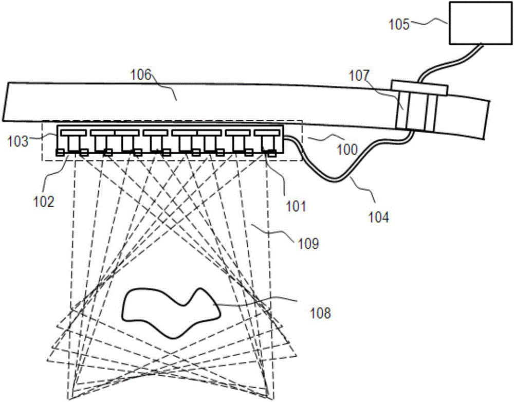
[0039] see figure 1 Shown is a structural view of the first embodiment of the three-dimensional endoscope of the present invention.
[0040] In this embodiment, the three-dimensional endoscope includes an imaging unit 100 and a control unit 105 .
[0041] The imaging unit 100 includes a housing 103 , and an imaging sensor array and an illumination device 101 located in the housing 103 . The imaging sensor array includes a plurality of imaging sensors 102 for acquiring a two-dimensional image of a target object 108 under illumination provided by an illumination device.
[0042] The control unit 105 is configured to synthesize a three-dimensional image of the target object based on the two-dimensional images of the target object 108 collected by each imaging sensor 102 .
[0043] In use, the imaging unit 100 may be placed inside the patient's body (eg, intraperitoneally), while the control unit 105 may be placed outside the patient's body.
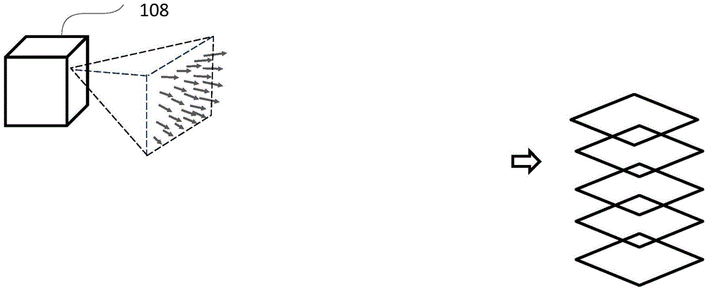
[0044] see figure 2 Shown is a s...
no. 2 approach
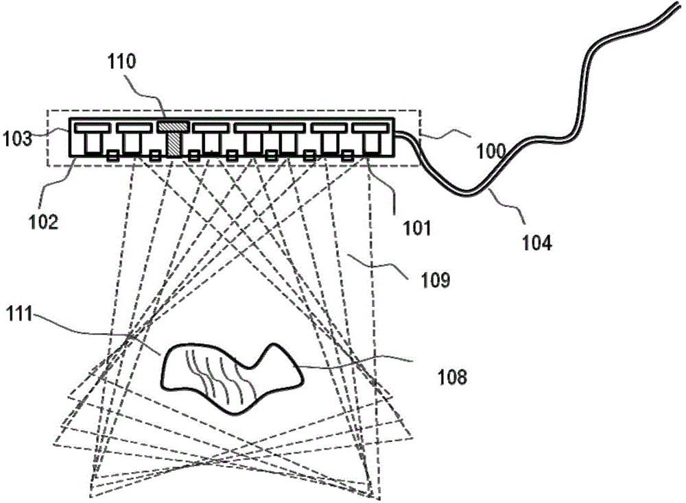
[0060] see image 3 Shown is a three-dimensional endoscope according to the second embodiment of the present invention.
[0061] In this embodiment, the lighting device 101 includes a structured light projection unit 110 .
[0062] The structured light projection unit 110 is used to generate a structured texture on the surface of the target object. Each imaging sensor 102 in the imaging sensor array is used to capture a two-dimensional image of the structured texture and transmit it to the control unit 105 . The control unit 105 performs three-dimensional reconstruction of the target object based on the plurality of two-dimensional images of structured textures.
[0063] exist image 3 , the structured light projection unit 110 generates a spatially varying structured texture 111 on the surface of the target 108 . Structured light is a well-known technique for imaging 3D surfaces. In the present invention, we apply the structured lighting technique to 3D endoscopy.
[00...
no. 3 approach
[0073] see Image 6 As shown, in this embodiment, the imaging sensor array includes a plurality of imaging sensors ( 302 - 305 ) with different spectral characteristics and polarization characteristics.
[0074] For multiple imaging sensors of the light field 3D endoscope, some sensors can be configured to acquire images in different wavelength bands and different polarization directions. For example, by adding a narrow-band filter, one or more imaging sensors can only capture light within a certain spectral range, thereby enhancing the imaging contrast (signal-to-noise ratio). Polarized imaging acquisition can suppress the impact of surface reflection on image quality.
[0075] Spectral imaging and polarization imaging are completely independent imaging methods. The two can be used simultaneously or independently according to specific application requirements.
[0076] Image 6 The illustrated imaging sensor array 301 has eight optical channels, each with its own unique s...
PUM
 Abstract
Description
Claims
Application Information
 - R&D Engineer
- R&D Manager
- IP Professional
- Industry Leading Data Capabilities
- Powerful AI technology
- Patent DNA Extraction
Browse by: Latest US Patents, China's latest patents, Technical Efficacy Thesaurus, Application Domain, Technology Topic.
© 2024 PatSnap. All rights reserved.Legal|Privacy policy|Modern Slavery Act Transparency Statement|Sitemap