Electrocardiogram signal R wave positioning method

An electrocardiographic signal and positioning method technology, applied in medical science, sensors, diagnostic recording/measurement, etc., can solve the problems of inaccurate preset thresholds, large amount of calculation, complicated methods, etc., and achieve fast and effective identification and simple calculation. , the effect of high accuracy

Active Publication Date: 2015-11-25
SHANGHAI NORMAL UNIVERSITY
View PDF11 Cites 3 Cited by
  • Summary
  • Abstract
  • Description
  • Claims
  • Application Information

AI Technical Summary

Benefits of technology

In this patented technology, an electrical cardiac activity (ECG) sensor uses specialized equipment that measures changes caused by heartbeats or bradyarrhythmia during physical exercise such as walking on uneven surfaces like grassy areas. By fitting specific curves with these differences from the measured data, it can accurately determine where certain parts of the patient's body have been affected without relying solely upon their own physician interpretation. These techniques make identifying any abnormalities easier than traditional methods involving measuring blood pressure levels alone.

Problems solved by technology

This patented technical problem addressed in this patents relates to improving the accuracy and efficiency of identifying specific types of electroencephalography (electrophysytics), particularly when analyzed over longer periods of data collection without recomputation capabilities.

Method used


Image

  • Electrocardiogram signal R wave positioning method
  • Electrocardiogram signal R wave positioning method
  • Electrocardiogram signal R wave positioning method

Examples


Embodiment Construction

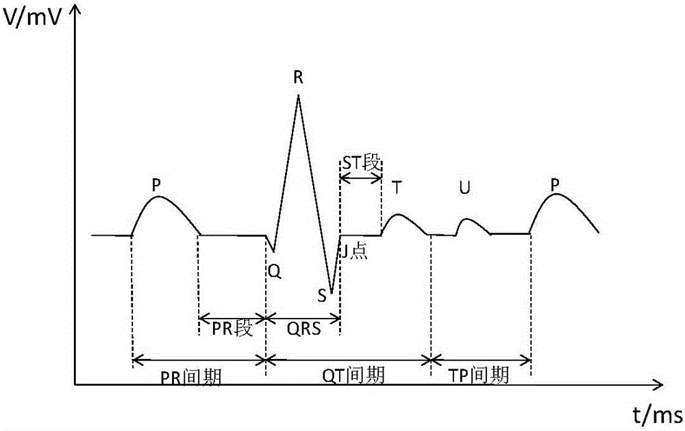
[0037] The human ECG signal reflects the regular depolarization and repolarization process of myocardial cells. The human heart signal S(t) is collected by the ECG detector, and the baseline drift, power frequency interference, and EMG interference are removed by filtering methods. Mixed interference signals, to obtain a clearer ECG signal Then, features such as QRS wave, RR interval, S wave, T wave, ST segment displacement, ST segment slope and other features of ECG signal are extracted by feature extraction method to reflect the strength of cardiac function.

[0038] The human ECG signal reflects the regular depolarization and repolarization process of myocardial cells. The human heart signal S(t) is collected by the ECG detector, and the baseline drift, power frequency interference, and EMG interference are removed by filtering methods. Mixed interference signals, to obtain a clearer ECG signal Then, features such as QRS wave, RR interval, S wave, T wave, ST segment disp...


PUM


Abstract

The invention discloses an electrocardiogram signal R wave positioning method, wherein, for filtered electrocardiogram signals, the method comprises the following steps: fitting the human electrocardiogram signals by virtue of an interpolation polynomial method, and enhancing a peak value of the electrocardiogram signals through double curve differentials, so as to determine the position of R wave. The method disclosed by the invention is not only simple in computation and easy in implementation, but also high in accuracy rate; therefore, a new way is provided for the rapid and effective recognition of the electrocardiogram signals.

Description


Claims


Application Information

Patent Timeline
Owner SHANGHAI NORMAL UNIVERSITY
Why Eureka
Try Eureka