Chromatography microscopic measurement method for parallel optical lines

A microscopic measurement and line layer technology, applied in measurement devices, optical devices, instruments, etc., can solve problems such as affecting measurement accuracy, limiting the application range of structured illumination obvious microtechnology, and reducing measurement efficiency.

Inactive Publication Date: 2016-04-06
XI AN JIAOTONG UNIV
View PDF5 Cites 0 Cited by
  • Summary
  • Abstract
  • Description
  • Claims
  • Application Information

AI Technical Summary

Problems solved by technology

[0004] In summary, the existing microscopic measurement technology generally uses mechanical step scanning, which not only reduces the measurement efficiency, but also affects the measurement accuracy. The above problems limit the application range of structured illumination microscopy technology.

Method used


Image

  • Chromatography microscopic measurement method for parallel optical lines
  • Chromatography microscopic measurement method for parallel optical lines
  • Chromatography microscopic measurement method for parallel optical lines

Examples


Embodiment

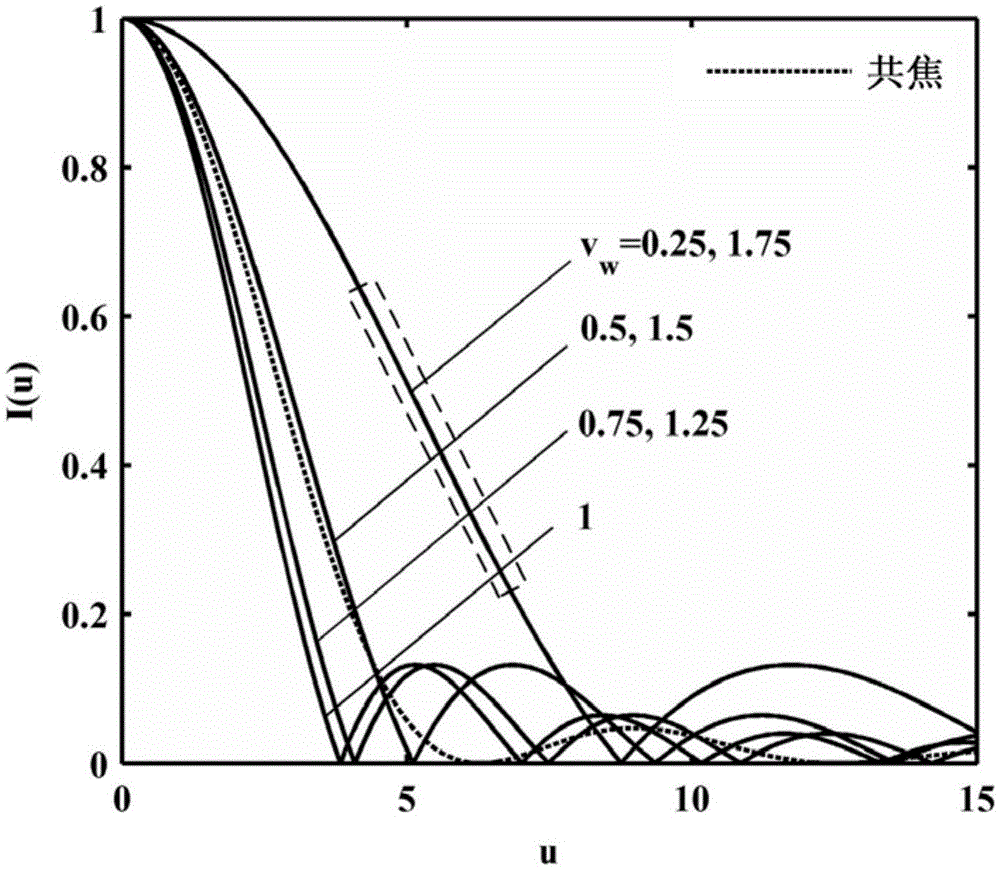
[0040] figure 2 Shown is the axial tomographic response curve of the structured light microscopy imaging system versus the normalized spatial frequency υ ω The law of change, when υ ω =1, ω=(sinα / λ) / M, the axial tomographic response curve is the narrowest, corresponding to the best axial resolution, that is, I(u)=|2J 1 (u) / u|, and the ideal confocal axial tomographic response I conf (u)=sinc 2 Compared with [u / (2π)] (shown by the dotted line in the figure), its full width at half maximum (FWHM) is narrower. Intercept the linear interval of the above curve (such as figure 2 As shown in the dotted line box), the theoretical correlation model between the intensity field and the sample surface height can be established, and a single scan can complete the detection of the three-dimensional tomographic structure of the sample surface morphology within a certain axial range.

[0041] According to the theoretical analysis of the present invention, design optical system paramete...


PUM


Abstract

The invention discloses a chromatography microscopic measurement method for parallel optical lines, and belongs to the technical field of optical microscopic imaging and precise measurement. The method comprises the steps: firstly calculating and obtaining a microscopic chromatography image field of a standard plane reflector through employing a formula shown in the description, wherein I1, I2 and I3 are respectively three structured light modulation fields; drawing an axial chromatography response intensity curve corresponding to the central point of the obtained microscopic chromatography image field; secondly cutting a single-side linear section of the curve, building a theoretical correlation mode of an intensity field and a sample surface height, obtaining a measurement calibration linear curve, and forming a height-intensity look-up table; and finally replacing the standard plane reflector with an actual to-be-measured sample, obtaining the surface height values of all points in a single scanning microscopic image field according to the height-intensity look-up table, and achieving the chromatography detection of the surface topography three-dimensional structure of the surface of the sample in a certain axial range. The method can be used for the non-axial mechanical scanning and parallel and three-dimensional chromatography quick detection.

Description

Technical field: [0001] The invention belongs to the technical field of optical microscopic imaging and precision measurement, and in particular relates to a parallel optical line tomographic microscopic measurement method. Background technique: [0002] Optical microscopic measurement devices have played an important role in promoting the process of human understanding and understanding of the microscopic scientific world. In the middle and late 1950s, M. Minsky, a postdoctoral fellow at Harvard University, invented a brand new optical microscopic device, that is, the common Focus microscope, which uses point illumination and point detection, and introduces a point-by-point scanning structure to realize three-dimensional imaging of the internal structure of the sample. In 1997, on the basis of long-term research and exploration in the field of confocal microscopy, M.A.A.Neil and T.Wilson of Oxford University in the United Kingdom proposed structured illumination microscopy ...

Claims


Application Information

Patent Timeline
Patent Type & Authority Applications(China)
IPC IPC(8): G01B11/25
Inventor 杨树明王通刘涛蒋庄德时新宇
Owner XI AN JIAOTONG UNIV
Why Eureka
Try Eureka