pet system with crystal or detector cell spacing

A radiation detector and crystal technology, applied in the field of positron emission tomography, can solve the problems of high cost of large FOV systems, and achieve the effect of increasing FOV and reducing costs

Active Publication Date: 2020-06-05
KONINKLJIJKE PHILIPS NV
View PDF5 Cites 0 Cited by
  • Summary
  • Abstract
  • Description
  • Claims
  • Application Information

AI Technical Summary

Problems solved by technology

The size of the field of view (FOV) that a PET system can cover is directly determined by the number of crystals used, making large FOV systems costly

Method used


Image

  • pet system with crystal or detector cell spacing
  • pet system with crystal or detector cell spacing
  • pet system with crystal or detector cell spacing

Examples


Embodiment Construction

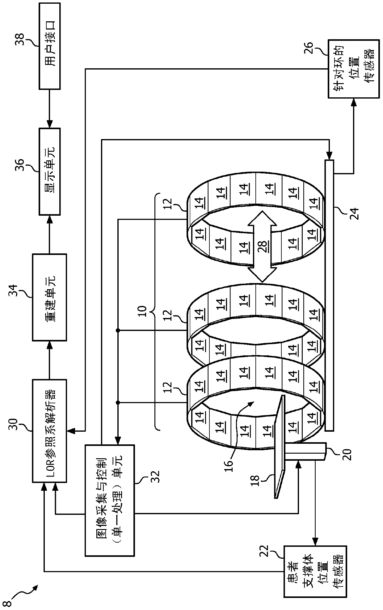
[0024] refer to figure 1 , imaging system 8 includes a positron emission tomography (PET) imaging system 10 and, optionally, an anatomical imaging system, such as a CT scanner (not shown). The PET scanner 10 includes a ring 12 of a plurality of detector units 14 housed within a gantry (not shown). The ring defines a patient receiving bore 16 . The imaging system 8 also includes a patient support 18, a patient support drive unit 20, and a position sensor 22 for the patient support unit. The ring 12 is movable by a ring positioner 24 (eg, motorized tracking, worm gear, etc.). Ring position sensor 26 outputs the position and rotational position of ring 12 to line of response (LOR) and frame of reference resolver 30 .

[0025] In a PET scan, a suitable positron-emitting radiopharmaceutical is administered to the subject prior to PET data acquisition. The emitted positrons undergo positron / electron annihilation, each annihilation event generating 511 keV gamma rays traveling in...


PUM


Abstract

A nuclear scanner comprising an annular support structure (12) supporting a plurality of radiation detector units (14), each detector unit comprising a crystal (52), a tile (66) containing an array of crystals ), or a module (14) of a tile. The detector cells define a circular array of crystals, and the circular array of crystals defines spaces between the columns. In another embodiment, the crystals define axial spaces between the crystals. The separated rings of crystals have staggered axial spaces so that the area of ​​the imaging field is not missed. The spaces between the detector units may be adjusted to form uniform or non-uniform spacing. Moving the patient through the annular support structure compensates for the reduced sampling in the space between arrays.

Description

technical field [0001] The present application relates to diagnostic imaging systems and methods. It finds particular application in positron emission tomography (PET), but also in other imaging systems. Background technique [0002] During a PET scan, the patient receives a dose of radiopharmaceutical. The drug is carried through the blood and accumulates and emits radiation in one or more target organs or areas. During a nuclear scanning procedure, emitted radiation is detected by the system and reconstructed into an image of the distribution of the radiopharmaceutical in the patient. The images can show the circulatory system and / or the relative uptake of the radiopharmaceutical in various regions or organs. For example, cancerous tumors take up significant amounts of glucose containing radiopharmaceuticals. The integration of the anatomical data from the anatomical scanning procedure and the metabolic data from the nuclear scanning procedure in the blended image prov...

Claims


Application Information

Patent Timeline
Patent Type & Authority Patents(China)
IPC IPC(8): A61B6/03A61B6/04A61B6/00G01T1/29
CPCA61B6/037A61B6/4258A61B6/4266A61B6/54G01T1/2985A61B6/0487A61B6/0407A61B6/4216A61B6/4275A61B6/4476A61B6/547
Inventor B·张C-H·童
Owner KONINKLJIJKE PHILIPS NV
Why Eureka
Try Eureka