
Multi-imaging modality navigation system

A navigation system and multi-modal technology, applied in image analysis, image data processing, medical science, etc., can solve problems such as increasing process time and patient discomfort

Active Publication Date: 2016-08-17
BK MEDICAL HLDG CO INC
View PDF7 Cites 4 Cited by
  • Summary
  • Abstract
  • Description
  • Claims
  • Application Information

AI Technical Summary

Problems solved by technology

This can lead to increased procedure time and patient discomfort

Method used


Image

  • Multi-imaging modality navigation system
  • Multi-imaging modality navigation system
  • Multi-imaging modality navigation system

Examples


Embodiment Construction

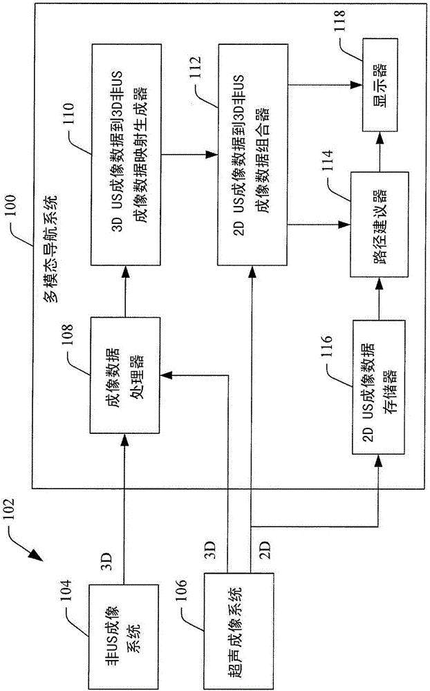
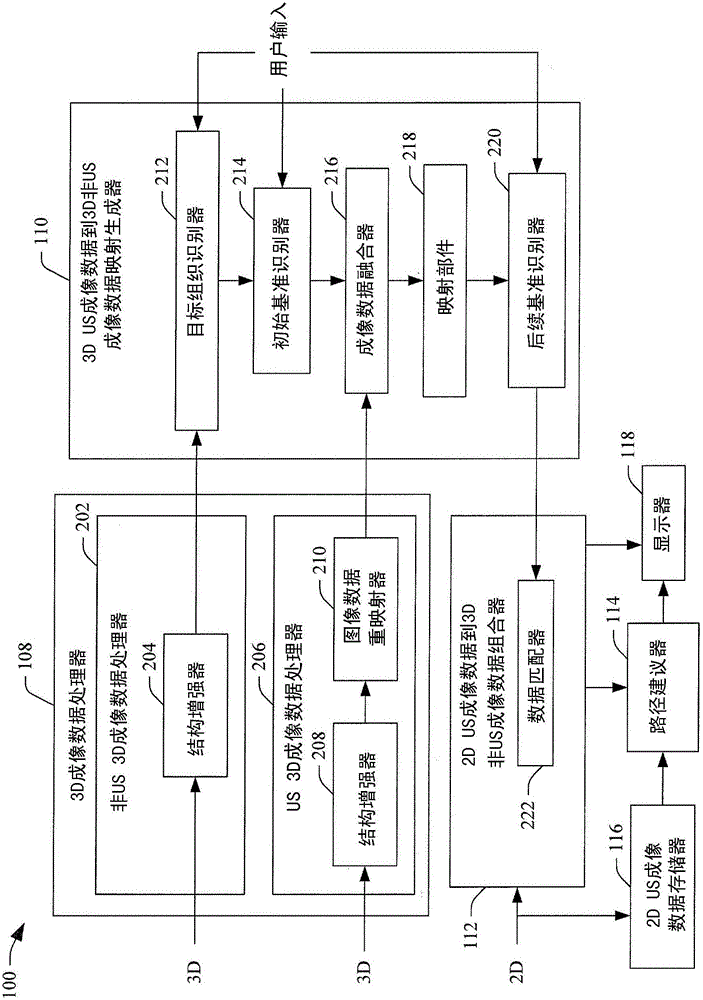
[0021] The following describes a method for visually presenting in both 2D imaging data and 3D imaging data during an imaging process of 2D imaging data acquired with a US imaging probe and previously acquired 3D imaging data of a volume of interest. A method to set fiducial markers to track the position of the US imaging probe relative to the volume of interest. Methods for suggesting movement of the probe to move the probe such that the target tissue of interest is in the field of view of the probe are also described below.

[0022] For example, application of the method in conjunction with, for example, a biopsy of the prostate gland facilitates localization of target tissue of interest. In one example, tracking and / or movement is achieved without using any external tracking systems (eg, electromechanical sensors) and / or segmenting tissue from 2D imaging data and / or 3D imaging data. As such, the methods described herein can ease hardware-based tracking, provide superior an...


PUM


Abstract

A method includes obtaining first 3D imaging data for a volume of interest. The first 3D imaging data includes structural imaging data and a target tissue of interest. The method further includes obtaining 2D imaging data. The 2D imaging data includes structural imaging data for a plane of the volume of interest. The plane includes at least three fiducial markers of a set of fiducial markers. The method further includes locating a plane, including location and orientation, in the first 3D imaging data that corresponds to the plane of the 2D imaging data by matching the at least three fiducial markers with corresponding fiducial markers identified in the first 3D imaging data and using the map. The method further includes visually displaying the first 3D imaging data with the 2D imaging data superimposed over at the corresponding plane located in the first 3D imaging data.

Description

technical field [0001] The following generally relates to imaging, and as such applies to multi-imaging modality navigation systems. Background technique [0002] Ultrasound imaging systems include transducer arrays that emit ultrasound beams into an examination field of view. As the beam penetrates structures in the field of view (e.g., of an object or a subsection of an object), subsections of the beam are attenuated, scattered, and / or reflected from the structure, with some reflections (echoes) throughout the return transduction device array. The transducer array receives the echoes, which are processed to generate an image of the object or a sub-portion of the object. The image is displayed visually. [0003] Ultrasound imaging is widely used in medical and non-medical applications. An example of a medical application is ultrasound guided biopsy. Generally, a biopsy is a procedure in which a small sample of tissue of interest (eg, prostate, lung, breast, etc.) is re...

Claims


Application Information

Patent Timeline
Patent Type & Authority Applications(China)
IPC IPC(8): G06T7/00
CPCA61B8/0841G06T7/33A61B8/085A61B8/483G06T7/37
Inventor N·A·阿科曼多D·利布利希J·P·奥康纳L·普洛
Owner BK MEDICAL HLDG CO INC
Learn More