CT image liver segmentation method and system based on automatic context model

A CT image and context technology, applied in the field of machine learning, can solve problems such as poor segmentation effect and difficult liver segmentation, and achieve the effect of improving segmentation results and improving segmentation accuracy

Active Publication Date: 2016-09-21
BEIJING INSTITUTE OF TECHNOLOGYGY
View PDF3 Cites 40 Cited by
  • Summary
  • Abstract
  • Description
  • Claims
  • Application Information

AI Technical Summary

Problems solved by technology

[0003] The existing random walk segmentation method has the advantages of being fast and simple, but it is not effective in segmenting areas with low contrast in CT images, especially the junctions between the liver and adjacent organs such as large blood vessels and stomach, which rely solely on the gray value Difficult to efficiently segment the liver

Method used


Image

  • CT image liver segmentation method and system based on automatic context model
  • CT image liver segmentation method and system based on automatic context model
  • CT image liver segmentation method and system based on automatic context model

Examples


Embodiment Construction

[0024] In order to make the purpose, technical solutions and advantages of the embodiments of the present invention clearer, the technical solutions in the embodiments of the present invention will be clearly described below in conjunction with the accompanying drawings in the embodiments of the present invention. Obviously, the described embodiments are the Some, but not all, embodiments are invented. Based on the embodiments of the present invention, all other embodiments obtained by persons of ordinary skill in the art without making creative efforts belong to the protection scope of the present invention.

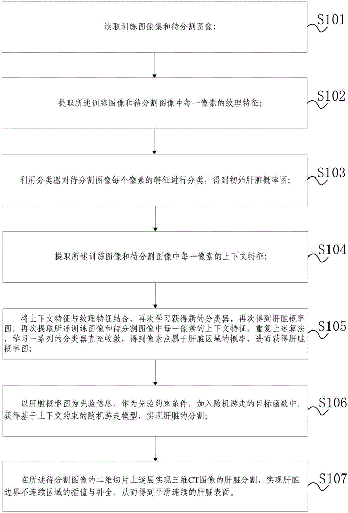
[0025] Such as figure 1 As shown, the present embodiment discloses a liver segmentation method for CT images based on an automatic context model, including:

[0026] S101. Read the training image set and the image to be segmented, wherein the training image in the training image set and the image to be segmented are CT images of the liver;

[0027] S102. Extracting te...


PUM


Abstract

The invention discloses a CT image liver segmentation method and system based on an automatic context model and capable of increasing liver segmentation precision in a CT image. The method comprises steps of: reading a training image set and a to-be-segmented image; extracting the textural feature of each pixel in the images; classifying the feature of each pixel in the to-be-segmented image by using a classifier to obtain an initial liver probability graph; extracting the context feature of each pixel in the images; combining the context features with the textural features and learning a series of classifiers by means of iteration until convergence to obtain a liver probability graph; using the liver probability graph as prior information and adding the liver probability graph a prior constraint condition to a randomly walking objective function as to obtain a random walk model based on context constraint for segmenting the liver; achieving liver segmentation of a three-dimensional CT image layer by layer on a two-dimensional slice of the to-be-segmented image, and interpolation and complement of a liver bound discontinuous area so as to obtain a smooth continuous liver surface.

Description

technical field [0001] The invention relates to the technical field of machine learning, in particular to a CT image liver segmentation method and system based on an automatic context model. Background technique [0002] Medical image segmentation assists doctors in identifying patients' internal tissues, organs and lesion areas, and plays a vital role in computer-aided treatment and surgical planning. Therefore, the automatic segmentation of the liver is the basis for doctors to diagnose and treat liver diseases such as cirrhosis, liver tumors, and liver transplantation. In abdominal CT images, the gray value difference between the liver and adjacent organs is small, and the liver itself has uneven gray levels and different shapes, so it is difficult to automatically and accurately segment the liver. Therefore, clinicians urgently need a simple, fast and accurate liver segmentation method. [0003] The existing random walk segmentation method has the advantages of being f...

Claims


Application Information

Patent Timeline
Patent Type & Authority Applications(China)
IPC IPC(8): G06T7/00G06T7/40G06K9/62
CPCG06T7/0012G06T2207/30056G06T2207/10081G06F18/241
Inventor 艾丹妮杨健王涌天丛伟建付天宇张盼王泽宇
Owner BEIJING INSTITUTE OF TECHNOLOGYGY
Why Eureka
Try Eureka