Method for analyzing histopathologic image and system thereof

A histopathology, image technology, applied in character and pattern recognition, instruments, computer parts, etc., can solve problems such as inability to extract local features

Active Publication Date: 2017-04-19
BEIJING CURACLOUD TECH CO LTD
View PDF6 Cites 18 Cited by
  • Summary
  • Abstract
  • Description
  • Claims
  • Application Information

AI Technical Summary

Problems solved by technology

However, the biggest defect of the SIFT feature is that it cannot accurately extract local features near the nucleus. Once the SIFT feature is extracted on the entire picture,

Method used


Image

  • Method for analyzing histopathologic image and system thereof
  • Method for analyzing histopathologic image and system thereof
  • Method for analyzing histopathologic image and system thereof

Examples


Embodiment Construction

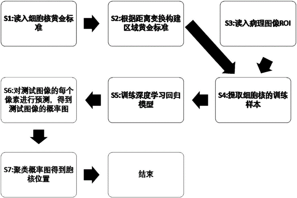
[0039] like figure 1 As shown, a method for detecting cell nuclei using a deep learning algorithm according to an embodiment of the present invention includes the following steps:

[0040] S1: Read in the gold standard of cell nuclei that is manually marked on histopathological images. The so-called gold standard of cell nuclei is the position of the cell nucleus that is manually marked, and only the position information of one pixel of the cell nucleus;

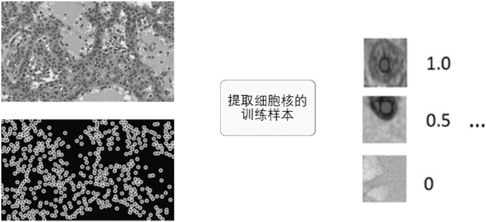
[0041] S2: According to the distance transformation, the regional gold standard is constructed in the histopathological image, so that each pixel near the nucleus gets a score to measure the distance from the pixel to the nucleus. The score falls in the range of 0-1, and the score at the center of the nucleus is 1, the farther away from the nucleus, the lower the score, and the background part is 0, such as figure 2 Among the extracted training samples shown, the score of the area where the top training sample is located i...


PUM


Abstract

The invention provides a method for analyzing a histopathologic image and a system thereof. The method comprises the steps that the central position of a cell nucleus is detected; then the graph structure features of the cell nucleus distribution are extracted; one or multiple graph structure features is/are calculated so that the global features of the cell nucleus distribution are obtained; the local features are directly extracted near the position of the cell nucleus; the global feature and the local features are fused so as to obtain the fused features; and the fused features are classified by using a classifier. The system comprises a high-resolution pathologic section scanner which scans the dyed histopathologic section to a computer so as to obtain a high-resolution digital histopathologic image; and the computer which performs the steps. The central position of the cell nucleus can be rapidly, accurately and automatically detected so that redundant information can be reduced, the overall distribution situation of the position of the cell nucleus and the detailed situation near the cell nucleus can be both considered, pathologic grading of the section can be accurately and rapidly realized and thus processing and transmission are rapid and resource consumption is low.

Description

technical field [0001] The invention relates to a computer-aided medical image processing technology, in particular to a method and system for analyzing histopathological images. Background technique [0002] Cancer is generally a heterogeneous disease with different risk grades, and its corresponding treatment options and prognosis vary from case to case. Taking the diagnosis of breast cancer as an example, mammography is usually used to determine the nature of the lesion first, and then the living tissue is obtained by puncture and stained to make a sample, and the stained sample is imaged to obtain a histopathological image. Most of the research on histopathological images focuses on the identification of specific tissue structures, such as cell nucleus detection, classification of malignant and benign pathological tissues, etc. The location, size, shape, and some specific structures of these tissues are very important indicators for disease diagnosis. The generation an...

Claims


Application Information

Patent Timeline
IPC IPC(8): G06K9/46G06K9/62
CPCG06V10/44G06V10/462G06F18/24
Inventor 王昕宋麒尹游兵曹坤林
Owner BEIJING CURACLOUD TECH CO LTD
Why Eureka
Try Eureka