
Intelligent identification method for epithelial cells in leucorrhea microscopic image

A technology for epithelial cells and microscopic images, applied in the field of medical digital image processing, can solve the problems of artificial recognition persistence, stability and objectivity, which are difficult to guarantee, doping, and low precision.

Inactive Publication Date: 2017-06-20
NINGBO MOSHI OPTOELECTRONICS TECH
View PDF5 Cites 26 Cited by
  • Summary
  • Abstract
  • Description
  • Claims
  • Application Information

AI Technical Summary

Problems solved by technology

The current detection method is to mix the leucorrhea with 0.9% NACL solution to make a glass slide, and the doctor observes it under a microscope. Because of the characteristics of various types of cells in the leucorrhea, complex components, intertwined cells, and difficult to distinguish the size of the area, this inspection method It is based on the experience and judgment of medical staff, mixed with many subjective factors, and at the same time, the efficiency is low and the accuracy is not high, which makes it difficult to guarantee the persistence, stability and objectivity of manual recognition.

Method used


Image

  • Intelligent identification method for epithelial cells in leucorrhea microscopic image
  • Intelligent identification method for epithelial cells in leucorrhea microscopic image
  • Intelligent identification method for epithelial cells in leucorrhea microscopic image

Examples


Embodiment Construction

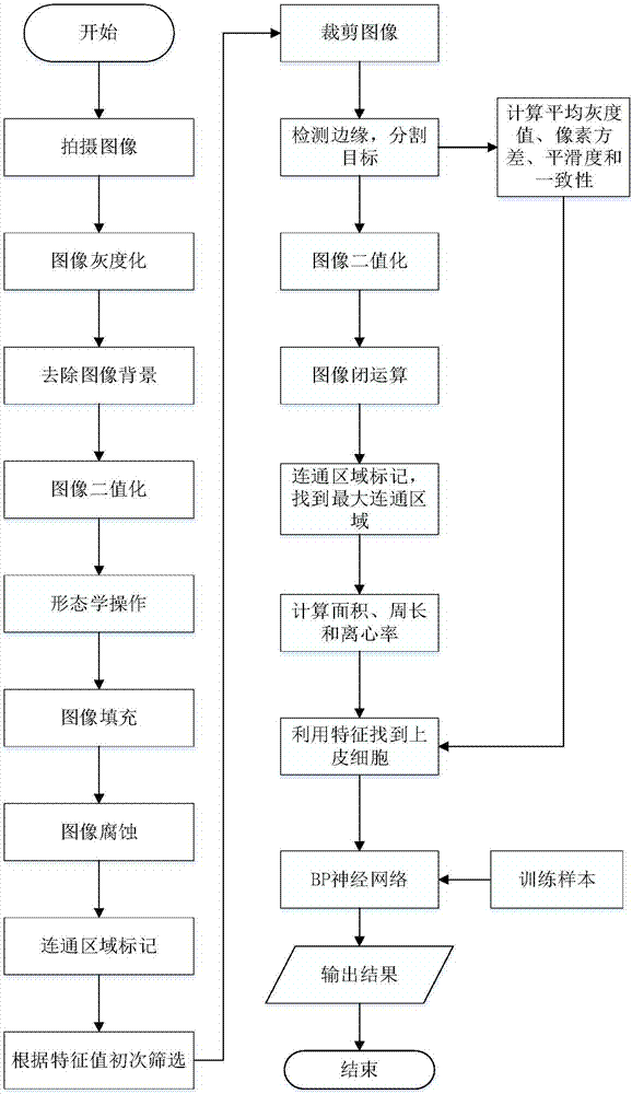
[0077] Below in conjunction with accompanying drawing, a kind of leucorrhea epithelial cell automatic detection method of the present invention is described in detail:

[0078] Step 1: Use a microscope to take images of leucorrhea mixed with 0.9% NACL solution to make a solution;

[0079] Step 2: Perform grayscale processing on the microscopic image taken in step 1 to obtain a grayscale image;

[0080] Step 3: remove the background of the grayscale image obtained in step 2;

[0081] Step 4: Binarize the image obtained in step 3;

[0082] Step 4-1: Perform morphological bottom-hat transformation on the image whose background has been removed to obtain a low-hat transformed image;

[0083] Step 4-2: use the grayscale threshold obtained by the maximum between-class variance method on the top-hat image;

[0084] Step 4-3: Compare the gray value of each pixel of the gray image with the gray threshold, if it is greater than the threshold, assign the gray value of 255 to the point...


PUM


Abstract

The invention provides an intelligent identification method for epithelial cells in a leucorrhea microscopic image. The method comprises the step that an image is photographed by a microscope; image processing is carried out, wherein the step of image processing comprises the steps of gray processing, binarization processing, morphological processing, filling and etching; communication area marking is carried out on the acquired image; the minimum value of the length and the width of the circumscribed rectangle of communication areas is greater than 85, and the maximum value is greater than 130 in communication areas are kept as the areas of suspected epithelial cells, wherein the area of the communication areas is greater than 4600 and the circumference is greater than 550; image processing is carried out on the suspected areas again; the eigenvalue of the maximum communication area is calculated and is compared with the eigenvalue of the epithelial cells; a BP neural network is input into a reserved area conforming to features; and epithelial cells are judged. According to the invention, a detection result can be accurately and quickly acquired; the influence of low efficiency and low precision of manual operation is eliminated; and the method has an important academic value and broad prospects for creating considerable social and economic benefits.

Description

technical field [0001] The invention belongs to the field of medical digital image processing, and specifically refers to an intelligent identification method for epithelial cells in leucorrhea microscopic images. Background technique [0002] Routine examination of leucorrhea is the most widely used examination for the diagnosis of gynecological diseases. The cleanliness of leucorrhea can be judged by observing the distribution of various cells in the microscopic image, so as to determine whether there is inflammation. Among them, the area occupied by epithelial cells is an important factor in determining the cleanliness of leucorrhea. The current detection method is to mix leucorrhea with 0.9% NACL solution to make a glass slide, and the doctor observes it under a microscope. Because of the characteristics of various types of cells in leucorrhea, complex components, intertwined cells, and difficult to distinguish the size of the area, this inspection method It is based on...

Claims


Application Information

Patent Timeline
IPC IPC(8): G06T7/11G06T7/187G06N3/06
CPCG06N3/061G06T2207/10056G06T2207/30004
Inventor 陈仕隆胡静蓉易少宾
Owner NINGBO MOSHI OPTOELECTRONICS TECH
Learn More