Determination method and system of salivary gland interesting areas

A technology for determining regions of interest and methods, which is applied in the field of medical diagnosis and can solve problems such as poor diagnostic accuracy

Inactive Publication Date: 2017-08-18
THE FIRST HOSPITAL OF CHINA MEDICIAL UNIV
View PDF3 Cites 3 Cited by
  • Summary
  • Abstract
  • Description
  • Claims
  • Application Information

AI Technical Summary

Problems solved by technology

[0005] Although the commonly used imaging examination of salivary gland dynamic imaging mentioned above can evaluate the function of salivary glands, the operation process of determining the location of the salivary gland region of interest mainly depends on the doctor's work experience, which is highly subjective and difficult to diagnose. poor

Method used


Image

  • Determination method and system of salivary gland interesting areas
  • Determination method and system of salivary gland interesting areas
  • Determination method and system of salivary gland interesting areas

Examples


Embodiment 1

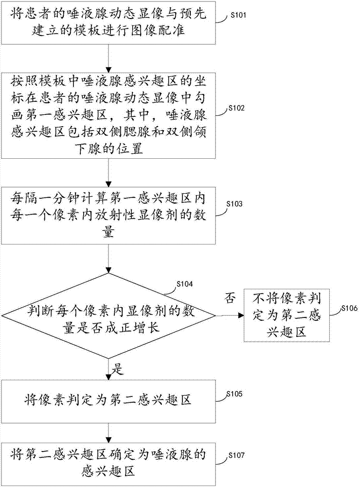
[0090] see figure 1 The method for determining the salivary gland region of interest proposed in this embodiment specifically includes the following steps:

[0091] Step S101: Register the patient's salivary gland dynamic imaging with a pre-established template. Here, the salivary gland dynamic imaging is an image taken 15 minutes after the patient is injected with a radioactive imaging agent. In order to facilitate the viewing of the results, radiopharmaceuticals are introduced into the patient's body before diagnosis, so that after the radiopharmaceuticals are introduced into the body, they can concentrate in the target organ or tissue, and the rays emitted by them are detected by imaging equipment, thereby obtaining The distribution images of drugs in the body are used as a basis to diagnose various diseases. There are many kinds of common radioactive imaging agents, for example, F-DOPA imaging agent, dopamine transporter imaging agent, dopamine receptor imaging agent, try...

Embodiment 2

[0123] see Figure 4 , the present embodiment provides a system for determining a salivary gland region of interest, including: a sequentially connected registration module 1, a first region of interest delineation module 2, a quantity statistics module 3, a first judgment module 4, an affirmative execution module 5, and a negative execution module Module 6 and ROI determination module 7, the specific functions of each module are introduced as follows:

[0124] The registration module 1 is used for image registration of the patient's salivary gland dynamic imaging with the pre-established template, and the first region of interest delineation module 2 is used for performing the image registration on the patient's salivary gland dynamic imaging according to the coordinates of the salivary gland region of interest in the template. Outline the first region of interest, wherein the salivary gland region of interest includes the positions of the bilateral parotid glands and bilater...


PUM


Abstract

The invention provides a determination method and system of salivary gland interesting areas, and relates to the technical field of medical diagnosis. The determination method of salivary gland interesting areas includes the steps: performing image registration on dynamic development of the salivary gland of a patient and a pre-established template; according to the coordinates of the salivary gland interesting areas in the template, sketching a first interesting area in the dynamic development of the salivary gland of the patient, wherein the salivary gland interesting areas include the positions of the parotid glands at two sides and submaxillary gland at two sides; every one minute, calculating the quantity of the radioactive developer in each pixel in the first interesting area; by means of the calculated quantity determining whether the quantity of the developer in each pixel has positive growth; if so, determining the pixel as the second interesting area; if not, not determining the pixel as the second interesting area; and finally, determining the second interesting area as the interesting area of the salivary gland. The determination method and system of salivary gland interesting areas can quickly and accurately sketch the interesting area of the salivary gland.

Description

technical field [0001] The invention relates to the field of medical diagnosis, in particular to a method and a system for determining a salivary gland region of interest. Background technique [0002] Salivary glands are glands in the oral cavity of humans or vertebrates that secrete saliva. The main function of the salivary glands is to secrete saliva, which is used to moisten the mouth on the one hand and facilitate swallowing and speaking on the other hand. Because the saliva secreted by the human body contains amylase, which can also initially decompose the starch in food. [0003] With the acceleration of the pace of life and the increase of people's life pressure, in recent years, the lesions of salivary glands have been increasing year by year, such as salivary gland inflammation, xerosis, swelling and other lesions, most of which will lead to salivary gland dysfunction. At present, the medical treatment method is to collect dynamic imaging of the patient's salivar...

Claims


Application Information

Patent Timeline
IPC IPC(8): G06K9/32G06T7/30
CPCG06T2207/10128G06T2207/30004G06V10/25
Inventor 陈松李亚明李雪娜尹雅芙李娜陈美洁刘光晖王姝
Owner THE FIRST HOSPITAL OF CHINA MEDICIAL UNIV
Why Eureka
Try Eureka