Medical imaging device and method for obtaining images for evaluating performance thereof

A technology of medical imaging and equipment, which is applied in the field of medical imaging, can solve the problems of inability to obtain the performance evaluation of complete rows of detectors, high cost of phantoms, and difficulty in meeting the requirements of the manufacturing process, so as to achieve fully automatic scanning and performance evaluation, saving Production cost and the effect of improving production efficiency

Active Publication Date: 2018-06-01
SHANGHAI UNITED IMAGING HEALTHCARE
View PDF11 Cites 4 Cited by
  • Summary
  • Abstract
  • Description
  • Claims
  • Application Information

AI Technical Summary

Problems solved by technology

Solve the problems in the performance evaluation method of multi-row CT imaging equipment in the prior art that the cost of the phantom is high, the manufacturing process is difficult to meet the requirements, and the performance evaluation of the complete number of detectors cannot be obtained

Method used


Image

  • Medical imaging device and method for obtaining images for evaluating performance thereof
  • Medical imaging device and method for obtaining images for evaluating performance thereof
  • Medical imaging device and method for obtaining images for evaluating performance thereof

Examples


Embodiment Construction

[0026] The present application will be further described below through specific embodiments and in conjunction with the accompanying drawings.

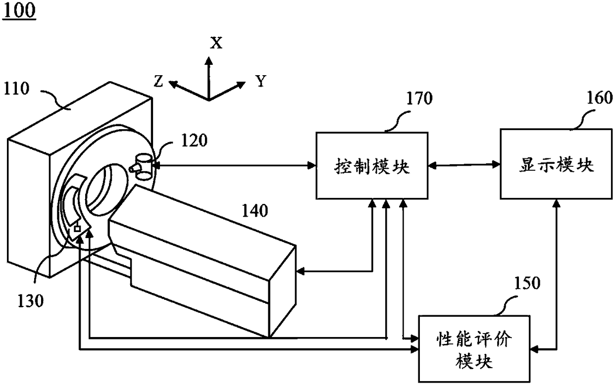
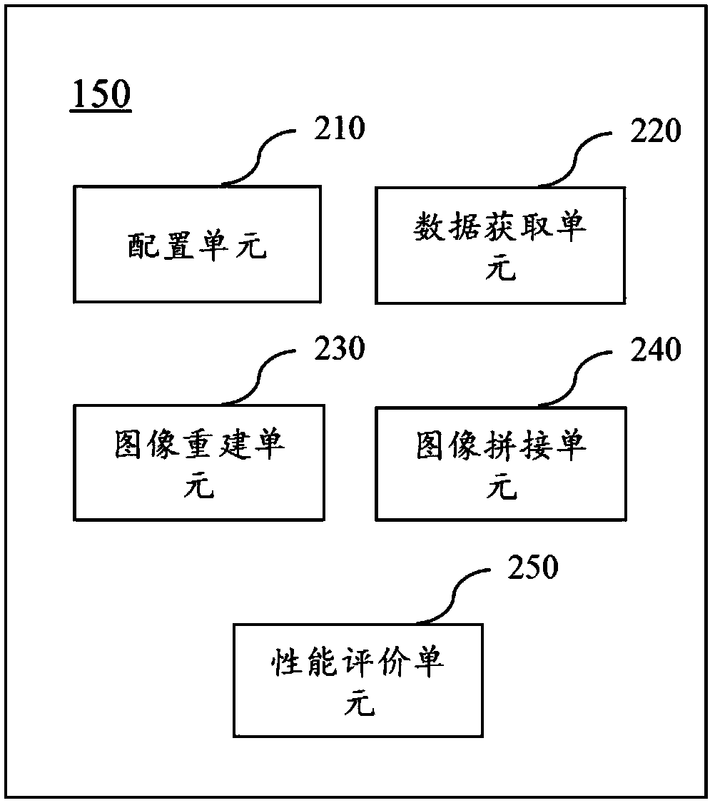
[0027] figure 1 is a schematic diagram of a medical imaging device according to some embodiments of the present application. Such as figure 1 As shown, the medical imaging device 100 may include a frame 110 , a radiation source 120 , a detection array 130 , a detection bed 140 , a scanning module 150 , a display module 160 and a control module 170 . The frame 110 may be used to support one or more components in the medical imaging device 100 . In some embodiments, a scanning cavity may be opened in the middle of the frame 110 . The ray source 120 may be used to emit rays or signals, and the rays may include X-rays, γ-rays and the like. The detection array 130 may be used to receive rays or signals after passing through the scanned object. Wherein, the detection array 130 may be disposed in the gantry 110 opposite to the radiation...


PUM


Abstract

The invention relates to a medical imaging device and a method for obtaining images for evaluating the performance thereof. The method comprises the steps of obtaining the effective thickness of a scanning object; obtaining the range of a detector of the medical imaging device; on the basis of the effective thickness of the scanning object and the range of the detector of the medical imaging device, automatically configuring the scanning mode of the medical imaging device; on the basis of the automatically-configured scanning mode, scanning he scanning object to obtain scanning data; on the basis of the scanning data, generating multiple scanning images; on the basis of the multiple scanning images, generating an integrated image, wherein the integrated image is used for evaluating the performance of the medical imaging device. The images used for evaluating the performance of the medical imaging device and obtained through the method do not depend on the die body size, the performanceof complete bay number detectors can be rapidly and conveniently evaluated, and the die body manufacturing cost is saved.

Description

technical field [0001] The present application relates to the field of medical imaging, in particular to a medical imaging device and a method for acquiring images used to evaluate the performance of the medical imaging device. Background technique [0002] In the field of medical diagnosis and treatment today, medical imaging equipment plays an important role, especially CT imaging equipment is one of the most important medical equipment at present. Before CT imaging equipment is used for human body scanning for medical diagnosis, the performance of CT imaging equipment needs to be evaluated in accordance with various regulations. The performance of CT imaging equipment mainly refers to the performance of the detector. Existing performance evaluation methods for CT imaging equipment rely on phantoms that comply with regulations. However, with the emergence of multi-row CT imaging equipment and the increase in the number of detector rows, the size of the phantom has to be i...

Claims


Application Information

Patent Timeline
Patent Type & Authority Applications(China)
IPC IPC(8): A61B6/00
CPCA61B6/52
Inventor 刘炎炎窦菲菲许鑫
Owner SHANGHAI UNITED IMAGING HEALTHCARE
Why Eureka
Try Eureka