CT image based automatic extraction method of 3D liver bounding volume

A CT image, automatic extraction technology, applied in the field of image processing, can solve the problems of high complexity, poor adaptability, sensitivity to noise, etc., to achieve the effect of improving efficiency, improving segmentation efficiency, and reducing data volume

Active Publication Date: 2018-07-06
NORTHEASTERN UNIV
View PDF8 Cites 6 Cited by
  • Summary
  • Abstract
  • Description
  • Claims
  • Application Information

AI Technical Summary

Problems solved by technology

These methods still have drawbacks such as: requiring manual interaction, sensitive to

Method used


Image

  • CT image based automatic extraction method of 3D liver bounding volume
  • CT image based automatic extraction method of 3D liver bounding volume
  • CT image based automatic extraction method of 3D liver bounding volume

Examples


Embodiment Construction

[0041] The present invention will be further elaborated below in conjunction with the accompanying drawings of the description.

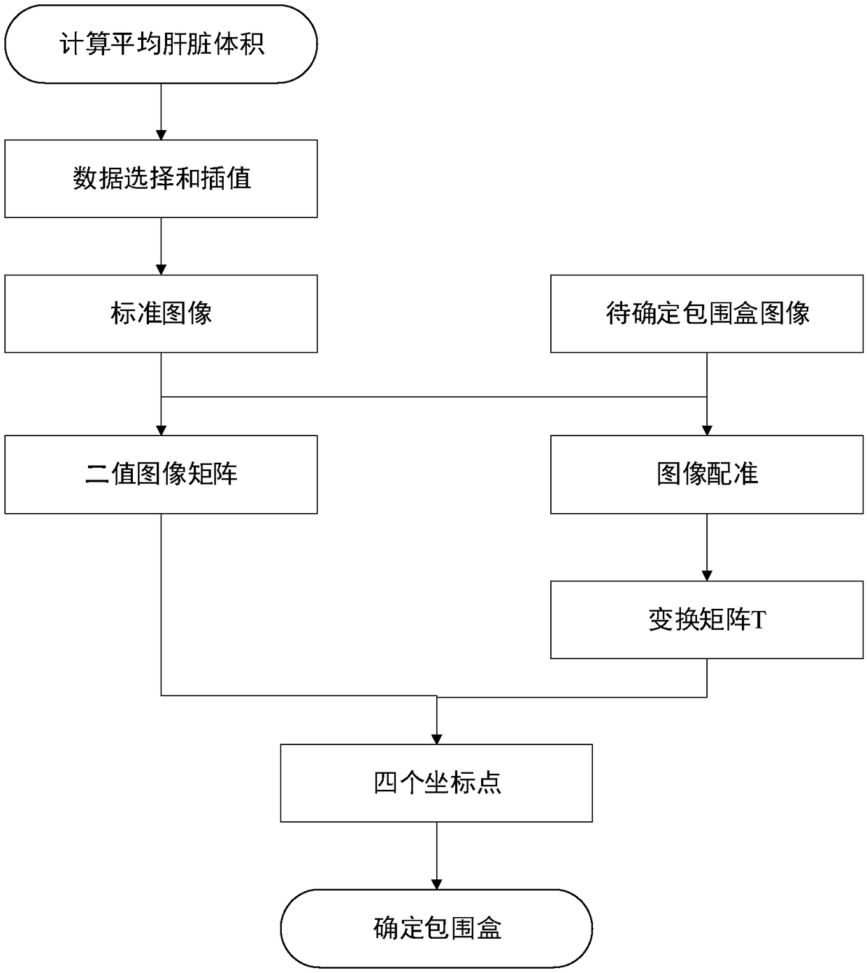
[0042] Such as figure 1 Shown, the present invention a kind of automatic extraction method of the three-dimensional liver bounding box based on CT image, comprises the following steps:

[0043] 1) Carry out volume measurement on abdominal CT liver segmentation data of N cases of people with different heights and weights, and calculate the average liver volume V a , and selected from these CT data and V a The closest three-dimensional CT slice and liver mask; in the present invention, N is 150-300.

[0044] 2) Interpolate the selected 3D CT slice and liver mask data along the vertical direction of the coronal plane;

[0045] 3) According to the liver mask data after interpolation, determine the liver area rectangle L of the corresponding CT slice r ;Complete the determination of the rectangular frame of the liver area for each CT slice after inte...


PUM


Abstract

The invention relates to a CT image based automatic extraction method of a 3D liver bounding volume. Volume measurement is carried out on liver segmentation data of N abdominal CT, the 3D CT slice anda liver mask layer in which the volume approaches the average liver volume most are selected, and a rectangular liver area frame corresponding to the CT slice is determined; a CT image of the determined rectangular liver area as a standard CT image is registered with a CT image of a non-determined liver bounding box roughly, the step and iteration frequency of an optimizer in rough registering are changed, and a transformation matrix is output; and geometric transformation is carried out on a binary image of the rectangular liver area frame, the matrix after transformation is placed in a 2D coordinate system, coordinates of all points whose grayscale values equal 1 are searched from the matrix, and a rectangular boundary is determined according to X and Y maximums and X and Y minimums, and serves as a liver area rectangle. The data bulk of image processing is reduced, rapid and accurate liver segmentation is guided, and the method has great significance in guiding segmentation of a liver tumor and provides basis for subsequent researches.

Description

technical field [0001] The invention relates to an image processing technology, in particular to an automatic extraction method of a three-dimensional liver bounding box based on a CT image. Background technique [0002] With the rapid development of science and technology and the continuous improvement of people's living standards, people pay more and more attention to their own health. Early and accurate detection of diseases and timely treatment can not only save the lives of patients, relieve their pain, but also reduce their pain. economic burden. Today, with the explosion of information technology, images, as the most basic, direct and real way for human beings to perceive the external environment, are constantly developing towards higher requirements. As a non-invasive medical diagnosis reference, medical imaging technology is playing an increasingly important role in modern medicine. Computed tomography (CT), ultrasound imaging (US), nuclear magnetic resonance (MRI...

Claims


Application Information

Patent Timeline
IPC IPC(8): G06T7/00G06T7/11G06T7/13G06T3/00G06K9/32
CPCG06T3/0075G06T7/0012G06T7/11G06T7/13G06T2207/10081G06T2207/30056G06V10/25
Inventor 姜慧研李少杰
Owner NORTHEASTERN UNIV
Why Eureka
Try Eureka