Vascular image reconstruction method and reconstruction device

A blood vessel image and blood vessel technology, which is applied in the field of computer-aided preoperative planning, can solve the problems of inaccurate and complete blood vessel tree images, high noise, and inability to satisfy the blood vessel tree images.

Active Publication Date: 2021-11-16
浙江京新术派医疗科技有限公司
View PDF3 Cites 0 Cited by
  • Summary
  • Abstract
  • Description
  • Claims
  • Application Information

AI Technical Summary

Problems solved by technology

In current CT imaging, contrast agents are used to enhance blood vessels, but the distribution of contrast agents in blood vessel images is not very ideal, and due to individual differences and the interference of various noises during imaging, the final image has low contrast. , characteristic of high noise
[0003] After using methods such as multi-scale vascular enhancement algorithm to segment some images with unsatisfactory quality (such as the images with high noise and low contrast characteristics mentioned above), the vascular image is segmented through the existing vascular image reconstruction method. The vascular image generated after vascular image reconstruction is not coherent, and the final vascular tree image is not accurate and complete enough, and there will still be a lot of noise interference
Existing vascular image reconstruction techniques cannot meet the actual needs of quickly and accurately generating complete vascular tree images in the process of vascular image reconstruction

Method used


Image

  • Vascular image reconstruction method and reconstruction device
  • Vascular image reconstruction method and reconstruction device
  • Vascular image reconstruction method and reconstruction device

Examples


Embodiment 1

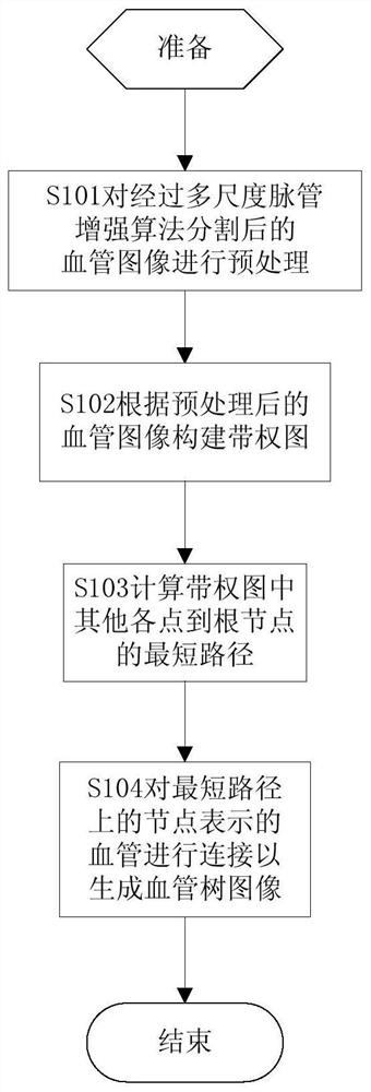
[0094] like Figure 1a As shown, in the first embodiment of the present invention, the vascular image segmented by the multi-scale vascular enhancement algorithm is preprocessed, a weighted graph is generated according to the preprocessed vascular image, and other nodes in the weighted graph are calculated The shortest path to the root node, and then connect the blood vessels represented by the nodes on the path according to the shortest path to generate the final vascular tree image.

[0095] S101. Preprocessing the blood vessel image segmented by the multi-scale vessel enhancement algorithm.

[0096] In the current CT image, since the gray value of the blood vessel is very close to the surrounding tissue, noise and interference will be generated during the imaging process, especially for some relatively small blood vessels, even if the contrast agent has been used in the CT image. Enhanced, the effect is very limited. Therefore, the multi-scale vessel enhancement algorithm ...

Embodiment 2

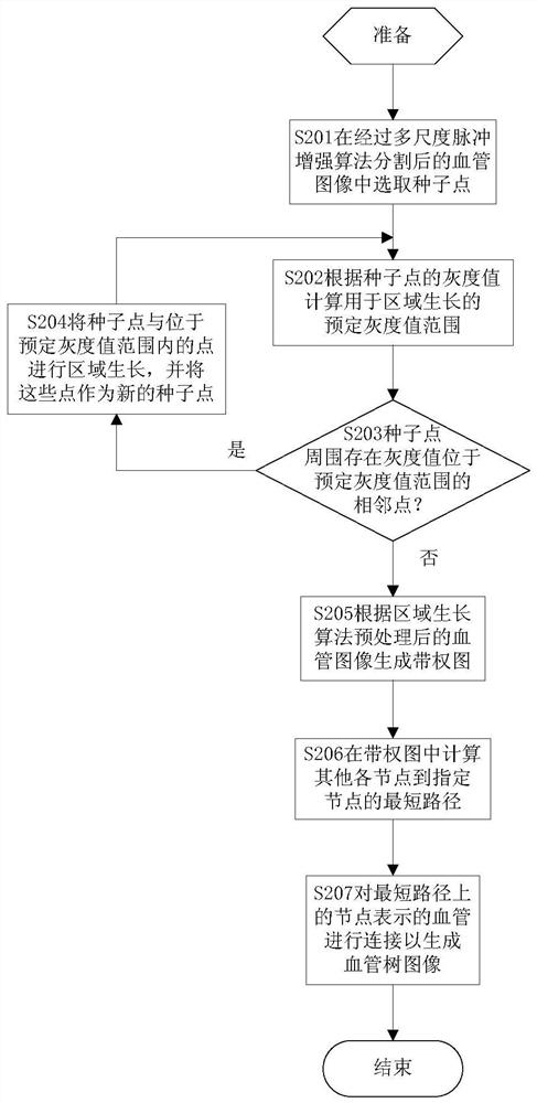
[0112] like Figure 2a As shown, in the second embodiment of the present invention, the region growing algorithm is used to preprocess the vascular image segmented by the multi-scale vascular enhancement algorithm, a weighted map is generated based on the preprocessed vascular image, and the weighted The shortest path from other nodes in the figure to the root node is used to connect the blood vessels represented by the nodes on the path according to the shortest path to obtain the reconstructed blood vessel tree image.

[0113] S201. Select seed points in the blood vessel image segmented by the multi-scale pulse enhancement algorithm.

[0114] The region growing algorithm connects the regions with similar gray values ​​near the seed point. This is an iterative process, and iterative growth is performed based on each seed point pixel.

[0115] In this embodiment, firstly, the blood vessels are segmented through the multi-scale vessel enhancement algorithm, and then the seed ...

Embodiment 3

[0131] like Figure 3a As shown, in the third embodiment of the present invention, the region growing algorithm is first used to perform the first preprocessing on the vessel image segmented by the multi-scale vessel enhancement algorithm, and then the directional expansion is used to perform the first preprocessing Carry out the second preprocessing of the blood vessel image, generate a weighted graph on this basis, calculate the shortest path from other nodes in the weighted graph to the root node, connect the blood vessels represented by the nodes on this path according to the shortest path, and obtain reconstruction image of the vascular tree.

[0132] S301. Perform first preprocessing on the blood vessel image segmented by the multi-scale vessel enhancement algorithm using a region growing algorithm.

[0133] For the principle of the multi-scale vascular enhancement algorithm, please refer to the description in step S101 of the first embodiment of the present invention. ...


PUM


Abstract

Embodiments of the present invention provide a method and device for reconstructing a blood vessel image. The method includes: preprocessing the blood vessel image segmented by a multi-scale vessel enhancement algorithm; A weighted graph of paths between nodes and related nodes, each path having a weight representing the distance between corresponding blood vessels; calculating the shortest path from other nodes in the weighted graph to a specified node; along the shortest path Different vessels represented by different nodes are connected to obtain a reconstructed vessel tree image. The vascular images segmented by the multi-scale vascular enhancement algorithm are preprocessed by using the region growing algorithm and the directional expansion algorithm. The device includes: a preprocessing module, a shortest path calculation module, and a blood vessel tree image generation module. The technical scheme provided by the invention can obtain more accurate and complete blood vessel tree images at a faster speed when the blood vessel image is reconstructed.

Description

technical field [0001] The present invention relates to computer technology, in particular to computer-aided preoperative planning technology. Background technique [0002] In the traditional preoperative planning process, the doctor observes 2D CT images, judges the positional relationship between the lesion and blood vessels based on experience and imagination, analyzes the difficulty and feasibility of the operation, and formulates a surgical plan accordingly. In current CT imaging, contrast agents are used to enhance blood vessels, but the distribution of contrast agents in blood vessel images is not ideal, and due to individual differences and the interference of various noises during imaging, the final image has low contrast. , high noise characteristics. [0003] After using methods such as multi-scale vascular enhancement algorithm to segment some images with less than ideal quality (such as the images with high noise and low contrast characteristics mentioned above...

Claims


Application Information

Patent Timeline
Patent Type & Authority Patents(China)
IPC IPC(8): G06T3/40G06T5/00G06T7/10
CPCG06T3/4038G06T5/002G06T2207/10081G06T2207/20024G06T2207/30101
Inventor 谢卫国王磊徐宁周龙宋晓堃
Owner 浙江京新术派医疗科技有限公司
Why Eureka
Try Eureka