Retinal vessel three-dimensional reconstruction method based on portable digital fundus camera

A retinal blood vessel and three-dimensional reconstruction technology, applied in ophthalmoscopes, eye testing equipment, 3D modeling, etc., can solve problems such as inability to reproduce fundus disease changes, little clinical significance, and inability to truly reconstruct the fundus

Active Publication Date: 2018-10-09
NANTONG UNIVERSITY
View PDF6 Cites 12 Cited by
  • Summary
  • Abstract
  • Description
  • Claims
  • Application Information

AI Technical Summary

Problems solved by technology

This method has a certain reference value for back projection reconstruction, but it only uses intermediate geometric pixels to represent the thickness and direction of bl

Method used


Image

  • Retinal vessel three-dimensional reconstruction method based on portable digital fundus camera
  • Retinal vessel three-dimensional reconstruction method based on portable digital fundus camera
  • Retinal vessel three-dimensional reconstruction method based on portable digital fundus camera

Examples


Embodiment Construction

[0033] In order to make the purpose, technical solutions and advantages of the embodiments of the present invention clearer, the technical solutions in the embodiments of the present invention will be clearly and completely described below in conjunction with the drawings in the embodiments of the present invention. Obviously, the described embodiments It is a part of embodiments of the present invention, but not all embodiments. Based on the embodiments of the present invention, all other embodiments obtained by persons of ordinary skill in the art without making creative efforts belong to the protection scope of the present invention.

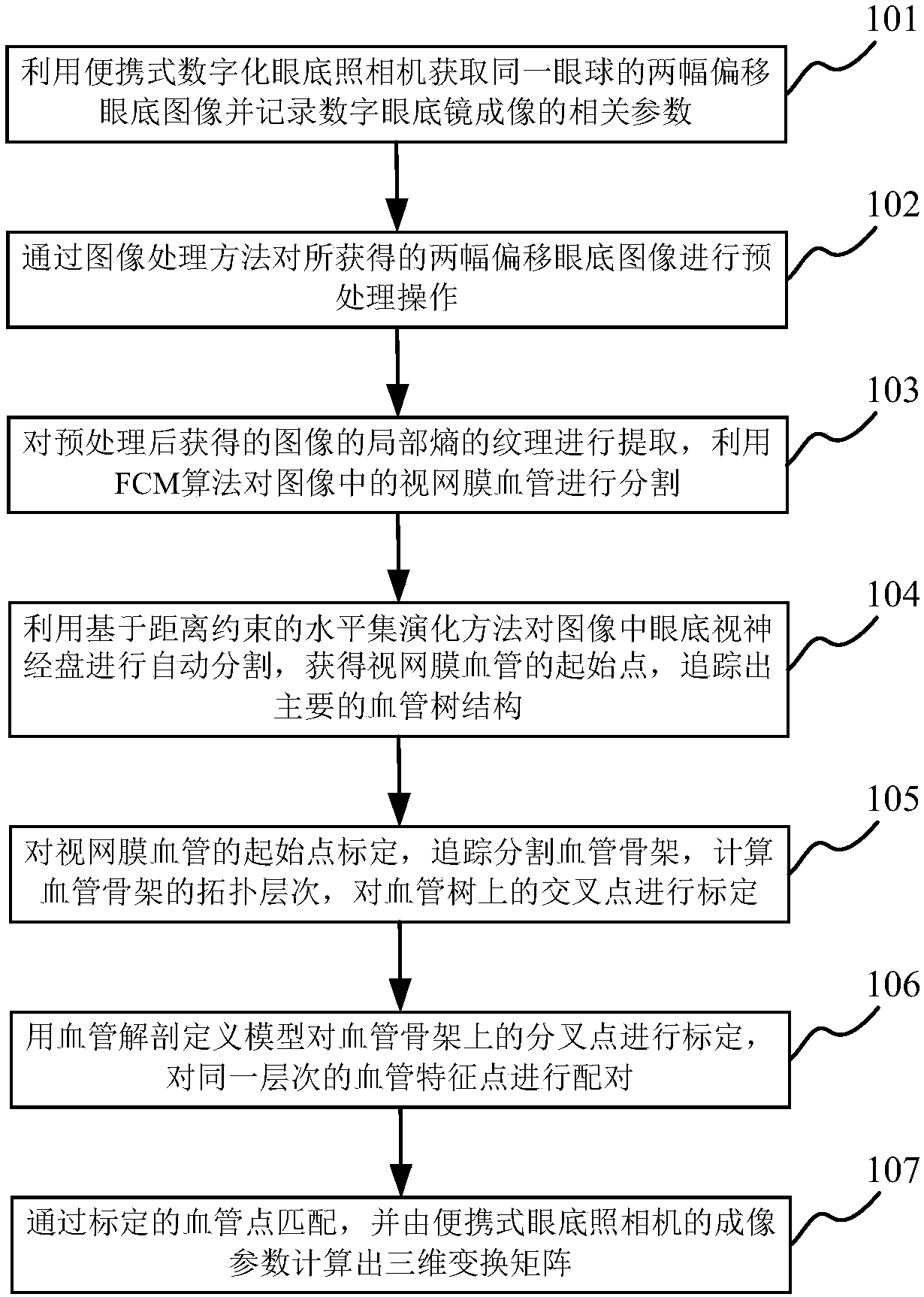
[0034] The present invention combines the retinal blood vessels of the same subject to have the same anatomical structure characteristics, and does not adopt the traditional calculation method based on image corner extraction and polar line constraints to obtain matching feature points, but proposes a method based on fundus blood vessel anatom...


PUM


Abstract

The invention belongs to the field of medical image processing and application, and provides a retinal vessel three-dimensional reconstruction method based on a portable digital fundus camera. The method comprise steps that firstly, a portable digital fundus image is used, the camera is used for two fundus images by camera displacement, both images are require to contain retinal optic nerve disc structures, and a certain amount of offset is generated; and then, the retinal vessel is segmented, and the feature points are extracted, and based on retinal vessel anatomical hierarchy characteristicpoint matching and a three-dimensional vessel reconstruction model, the retinal vessel is subjected to three-dimensional reconstruction. The retinal vessel three-dimensional reconstruction method based on the portable digital fundus camera can provide new supplement to an existing fundus blood vessel observation and analysis based on two-dimension.

Description

technical field [0001] The invention relates to the field of medical image processing and application, in particular to a three-dimensional retinal vessel reconstruction method based on a portable digital fundus camera. Background technique [0002] Fundus retinal microvessels are part of the vascular circulation system of the human body. Since they can be directly observed through the ophthalmoscope, they are of great significance for the early diagnosis and prognosis of many clinical diseases that affect the cardiovascular system, such as diabetes and hypertension. In the traditional fundus examination mode, the ophthalmologist directly inspects the fundus after the patient's pupil is dilated, but the changes of the fundus cannot be effectively recorded. With the continuous development of digital technology in recent years, digital photography can conveniently acquire, save and transmit images. Because digital fundus cameras can digitally collect fundus images, they are w...

Claims


Application Information

Patent Timeline
IPC IPC(8): A61B3/12A61B3/14A61B3/00G06T5/10G06T5/00G06T17/00
CPCA61B3/0025A61B3/12A61B3/14A61B2560/0431G06T5/007G06T5/10G06T7/0012G06T17/00G06T2207/20064G06T2207/30041G06T2207/30101G06T2210/41
Inventor 吴辉群张晓峰董建成裘茗烟郏科人吴英成
Owner NANTONG UNIVERSITY
Why Eureka
Try Eureka