Cell smear image acquisition and analysis system

A cell smear and image acquisition technology, which is applied in the analysis of materials, material analysis through optical means, and measurement devices, can solve problems that affect the efficiency of detection and the accuracy of results

Active Publication Date: 2018-11-30
深圳辉煌耀强科技有限公司
View PDF13 Cites 12 Cited by
  • Summary
  • Abstract
  • Description
  • Claims
  • Application Information

AI Technical Summary

Problems solved by technology

Although there are automated processes for sampling, smear preparation or cell identification in the prior art, the actual judgment still requires the participation of medical personnel, for example, the control of the stage, the judgment of cell identification, the preparation of analysis reports, etc., due to The difference in the level of medical personnel affects the efficiency of testing and the accuracy of results

Method used


Image

  • Cell smear image acquisition and analysis system
  • Cell smear image acquisition and analysis system
  • Cell smear image acquisition and analysis system

Examples


Embodiment Construction

[0051] According to the following detailed description of specific embodiments of the application in conjunction with the accompanying drawings, those skilled in the art will be more aware of the above and other objectives, advantages and features of the application.

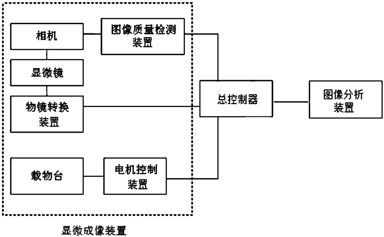
[0052] The embodiment of the present application provides a system for acquiring and analyzing cell smear images. figure 1 It is a schematic structure diagram of the cell smear image acquisition and analysis system according to the present application. The system includes: a stage for carrying cell smears; a motor control device connected to the stage for controlling the movement of the stage in three-dimensional space; an objective lens conversion device; a microscope connected to the stage The objective lens conversion device is connected to observe the cell smear; the camera is connected to the microscope to take images observed by the microscope; the image quality detection device is used to connect to the c...


PUM


Abstract

The application discloses a cell smear image acquisition and analysis system. The system comprises an objective table, a motor control device, an objective lens conversion device, a microscope, a camera, an image quality detection device, a main controller and an image analysis device, wherein the objective table is used for carrying a cell smear; the motor control device is connected with the objective table and is used for controlling the movement of the objective table; the microscope is connected with the objective lens conversion device; the camera is connected with the microscope and isused for shooting images observed by the microscope; the image quality detection device is connected with the camera; the main controller is separately connected with the image quality detection device, the objective lens conversion device and the motor control device; the image analysis device is connected with the controller. The system can automatically take pictures of the cell smear, analyzesthe image quality, and adjusts the light, thus obtaining clear images; a complete picture of the cell smear can be obtained by means of image stitching, so that the image can be analyzed more completely and comprehensively.

Description

technical field [0001] The present application relates to the technical field of automatic processing, in particular to a cell smear image acquisition and analysis system. Background technique [0002] In order to diagnose whether a patient suffers from or is predisposed to cancer, a cell sample needs to be removed from the patient and thoroughly analyzed in order to evaluate whether abnormal or abnormal cells are present. A pathologist or other skilled medical practitioner makes a diagnosis based primarily on specific characteristics of the cells in the sample. These cytological assays are based on the two-dimensional representation of the cells present in the sample, and most require immobilization of the cells on a matrix and the use of dyes or stains to visualize specific features of the cells. Although there are automated processes for sampling, smear preparation or cell identification in the prior art, the actual judgment still requires the participation of medical pe...

Claims


Application Information

Patent Timeline
Patent Type & Authority Applications(China)
IPC IPC(8): G01N21/84G01N21/01
CPCG01N21/01G01N21/84G01N2021/0112
Inventor 郏东耀李玉娟曾强
Owner 深圳辉煌耀强科技有限公司
Why Eureka
Try Eureka