Automatic segmentation method of brain tumor images based on convolutional neural network

A convolutional neural network and automatic segmentation technology, applied in the field of medical image segmentation and deep learning, can solve the problems of long image preprocessing time, rough segmentation of brain tumor images, and unbalanced categories, so as to shorten the processing time and improve the performance. The effect of identifying and resolving class imbalance

Active Publication Date: 2021-10-15
UNIV OF ELECTRONICS SCI & TECH OF CHINA
View PDF3 Cites 0 Cited by
  • Summary
  • Abstract
  • Description
  • Claims
  • Application Information

AI Technical Summary

Problems solved by technology

[0005] The purpose of the present invention is to provide a method for automatic segmentation of brain tumor images based on convolutional neural networks, which solves the technical problems of rough segmentation of brain tumor images, unbalanced categories and long image preprocessing time in the prior art

Method used


Image

  • Automatic segmentation method of brain tumor images based on convolutional neural network
  • Automatic segmentation method of brain tumor images based on convolutional neural network
  • Automatic segmentation method of brain tumor images based on convolutional neural network

Examples


Embodiment 1

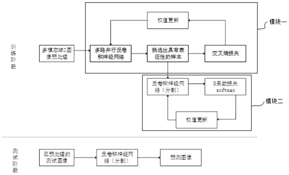
[0037] The method for automatically segmenting brain tumor images based on convolutional neural networks in the present invention includes multimodal MRI images of brain tumors, and further includes the following steps:

[0038] Step 1. Acquire multimodal MRI images of brain tumors and perform image preprocessing to obtain an original image set;

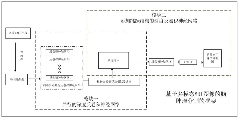
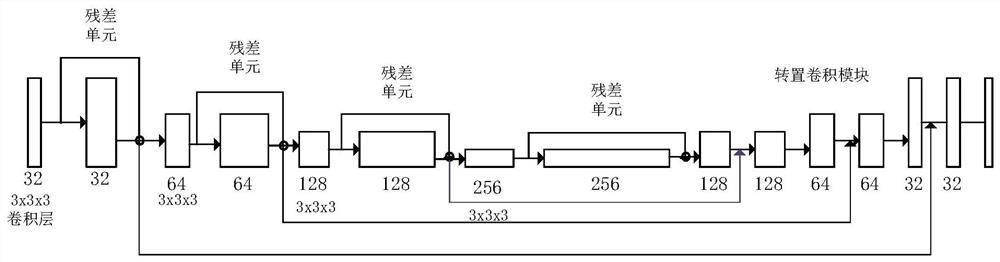
[0039] Step 2. Construct a framework for brain tumor segmentation based on multimodal MRI images; the framework includes module one and module two, and module one includes 3D convolutional neural network, residual unit and transposed convolution as the basis to form a parallel The deep deconvolution neural network is used to output the contour map of the brain tumor segmentation image; the second module includes adding a skip structure on the basis of the deep deconvolution neural network structure in the module one, which is used to output the lesion region segmentation of the brain tumor image picture;

[0040] Step 3. Input the o...

Embodiment 2

[0048] This embodiment makes the following further limitations on the basis of Embodiment 1: in the step 1, the multimodal MRI images are four kinds of modality images Flair, T1, T2, T1C, and the two modes of Flair and T2 N4ITK is used to correct the bias field of the images in the different modalities, and adjust the contrast of the images in the T1C and T1 modalities. Grayscale normalization of images between different individuals: first subtract the mean value of the entire image and divide by the standard deviation of the brain region, adjust the pixel values ​​​​of all images to the [-5, 5] interval, and normalize the entire image Normalized to [0, 1], non-brain regions are set to 0. Finally, translation transformation, twist enhancement and elastic deformation are performed on the preprocessed data. The detailed process is as follows: firstly, multimodal MRI images of brain tumors need to be acquired. In this method, only four modal images of Flair, T1, T2, and T1C are ...


PUM


Abstract

The invention discloses a brain tumor image automatic segmentation method based on a convolutional neural network, comprising the following steps: collecting multimodal MRI images of brain tumors and performing image preprocessing to obtain an original image set; constructing a brain tumor image based on multimodal MRI images. The framework of tumor segmentation; the framework includes module one and module two, module one includes a parallel depth deconvolution neural network based on 3d convolutional neural network, residual unit and transposed convolution; module two is included in the Based on the deep deconvolutional neural network structure in module 1 above, a skip structure is added; through several iterations of training, the ideal weight is obtained, and the segmentation map of the brain tumor image is output; the image segmentation results are tested and evaluated. The invention effectively solves the problem of low segmentation accuracy of brain tumor segmentation images; it can improve the identifiability of tumors and make image preprocessing operations more convenient; the use of the loss function based on the d coefficient in the segmentation module can effectively solve the category imbalance The problem.

Description

technical field [0001] The invention relates to the fields of medical image segmentation and deep learning, in particular to a method for automatic segmentation of brain tumor images based on convolutional neural networks. Background technique [0002] Medical image segmentation is a key technology in image analysis and processing. Separating relevant tissues of interest according to the similarity and specificity of the image area is of great significance to the clinical diagnosis and treatment process and is the main premise of all follow-up work. , the quality of the segmentation effect will directly affect the smooth progress of the information processing work. Medical image segmentation of brain tumors is an important branch in the field of image segmentation. Brain tumor segmentation technology plays an important role in the clinical diagnosis and treatment of brain tumors. Through the segmentation results of brain tumors, doctors can measure tumor Size and location, ...

Claims


Application Information

Patent Timeline
Patent Type & Authority Patents(China)
IPC IPC(8): G06T7/11G06T7/12G06N3/04
CPCG06T7/11G06T7/12G06T2207/30096G06T2207/30016G06T2207/20084G06T2207/20081G06T2207/10088G06N3/045
Inventor 程建郭桦苏炎洲高银星许轲
Owner UNIV OF ELECTRONICS SCI & TECH OF CHINA
Why Eureka
Try Eureka