
Method and device for processing three-dimensional medical model

A processing method and medical technology, applied in the field of medical imaging, can solve problems such as time-consuming and labor-intensive repairs of disconnected organs, and achieve the effects of convenient blood vessel completion and easy analysis of diseases

Pending Publication Date: 2019-01-18
QINGDAO HISENSE MEDICAL EQUIP
View PDF5 Cites 0 Cited by
  • Summary
  • Abstract
  • Description
  • Claims
  • Application Information

AI Technical Summary

Problems solved by technology

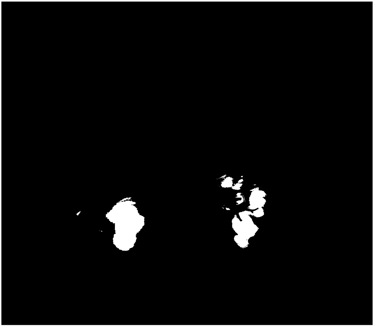
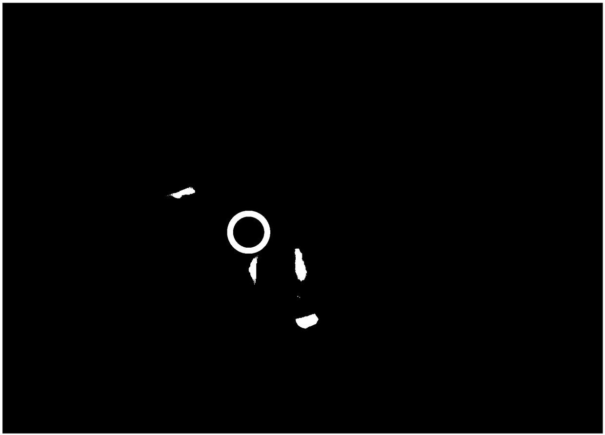
However, there are hundreds of medical images in the medical image sequence. It is time-consuming and labor-intensive to determine the image corresponding to the organ disconnection and the position of the organ disconnection in the image in the two-dimensional medical image sequence, which makes the organ disconnection Repairs are difficult to achieve

Method used


Image

  • Method and device for processing three-dimensional medical model
  • Method and device for processing three-dimensional medical model
  • Method and device for processing three-dimensional medical model

Examples


Embodiment Construction

[0051]Before explaining and describing the embodiments of the present invention in detail, the application scenarios of the embodiments of the present invention are firstly introduced. The method provided by the embodiment of the present invention is applied to a terminal. The terminal is a medical device in a medical scene. The medical device may be a display device of a medical image, such as a computer, a CT machine, an MRI instrument, etc., and the medical image may be a two-dimensional medical image. , a three-dimensional medical reconstruction model, etc., which are not limited in this embodiment of the present invention. As an example, the method provided by the embodiment of the present invention can also be applied to a computer-aided medical display device, which belongs to the field of computer-aided medical diagnosis. Computer Aided Diagnosis (CAD) refers to the use of imaging, medical image processing technology and other possible physiological and biochemical mea...


PUM


Abstract

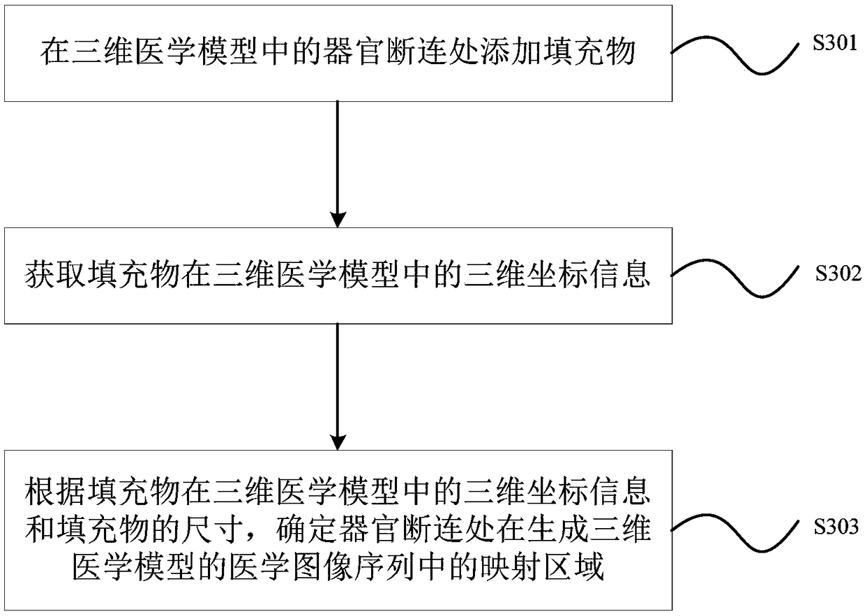
The invention provides a processing method and a device for a three-dimensional medical model. The method comprises the following steps: a filler is added to an organ disconnection part in the three-dimensional medical model; the filler is used for connecting the organ disconnection part; Obtaining the three-dimensional coordinate information of the filler in the three-dimensional medical model; According to the three-dimensional coordinate information of the filler in the three-dimensional medical model and the size of the filler, the mapping region of the organ disconnection in the medical image sequence that generates the three-dimensional medical model is determined. The processing method and the device of the three-dimensional medical model provided by the invention can quickly and accurately locate the organ disconnection place in the three-dimensional medical model.

Description

technical field [0001] The invention relates to medical image technology, in particular to a processing method and device for a three-dimensional medical model. Background technique [0002] With the development and application of medical imaging technologies such as computer graphics, computed tomography (Computed Tomography, CT), magnetic resonance imaging (Magnetic Resonance Imaging, MRI), ultrasound (ultrasonic, US) and other medical imaging technologies, traditional two-dimensional medical images can be reconstructed A three-dimensional medical model. The 3D medical model can clearly display the size and shape of human organs, blood vessels, lesions, and the positional relationship of surrounding tissues. It can assist doctors in diagnosis and treatment more vividly and three-dimensionally, and improve the accuracy and efficiency of diagnosis and treatment planning. [0003] The structure of human organs is complex, especially the blood vessels. The blood vessels in th...

Claims


Application Information

Patent Timeline
IPC IPC(8): G06T17/00G16H30/40
CPCG06T17/00G16H30/40G06T2210/41
Inventor 田广野王建军韩月乔
Owner QINGDAO HISENSE MEDICAL EQUIP
Learn More