Multi-scale nasopharyngeal tumor segmentation based on CNN

A multi-scale, nasopharyngeal technology, applied in image analysis, image data processing, instruments, etc., can solve problems such as lack of original feature information reuse, insufficient global feature information learning, and network inability to learn global features, and achieve good generalization ability. Effect

Inactive Publication Date: 2019-02-26
CHENGDU UNIV OF INFORMATION TECH
View PDF5 Cites 27 Cited by
  • Summary
  • Abstract
  • Description
  • Claims
  • Application Information

AI Technical Summary

Problems solved by technology

However, this approach will cause two problems: a large number of redundant calculations lead to low time efficiency, and the network cannot learn global features
However, this method does not extract enough global feature information, especially when the amount of data is small, it cannot fully learn global features.
In summary, there are two problems in the above method, on the one hand, lack of reuse of original feature information, on the other hand, insufficient learning of global feature information

Method used


Image

  • Multi-scale nasopharyngeal tumor segmentation based on CNN
  • Multi-scale nasopharyngeal tumor segmentation based on CNN
  • Multi-scale nasopharyngeal tumor segmentation based on CNN

Examples


Embodiment Construction

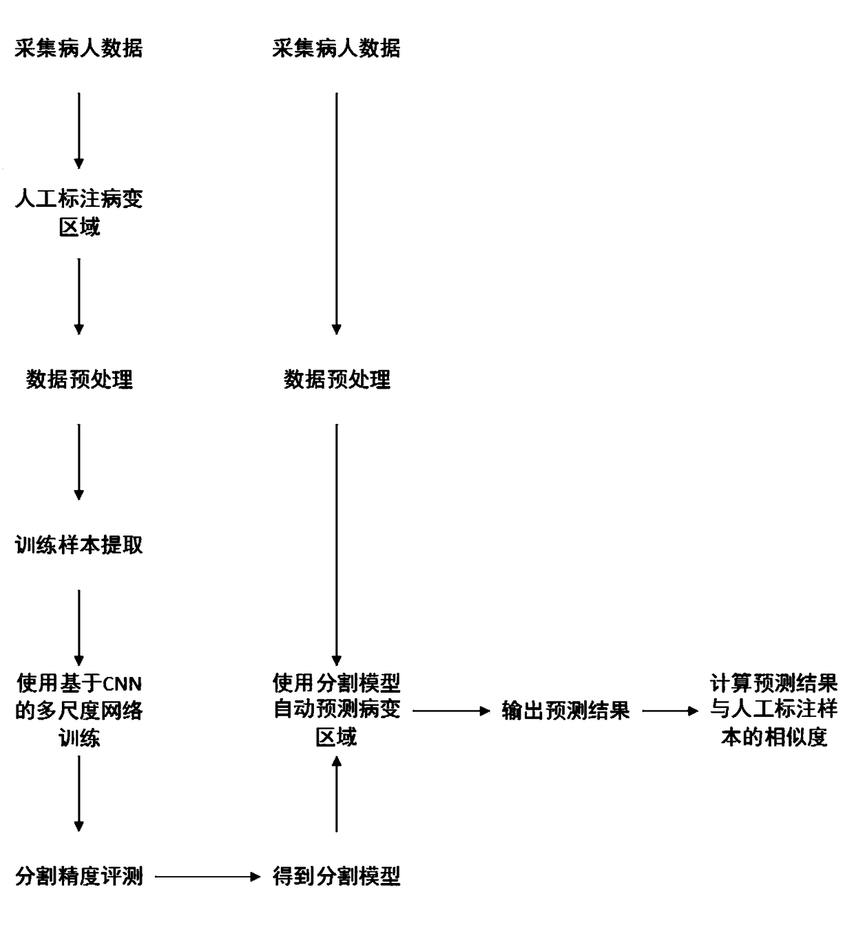
[0041] The present invention will be further described in detail below in conjunction with the accompanying drawings and embodiments.

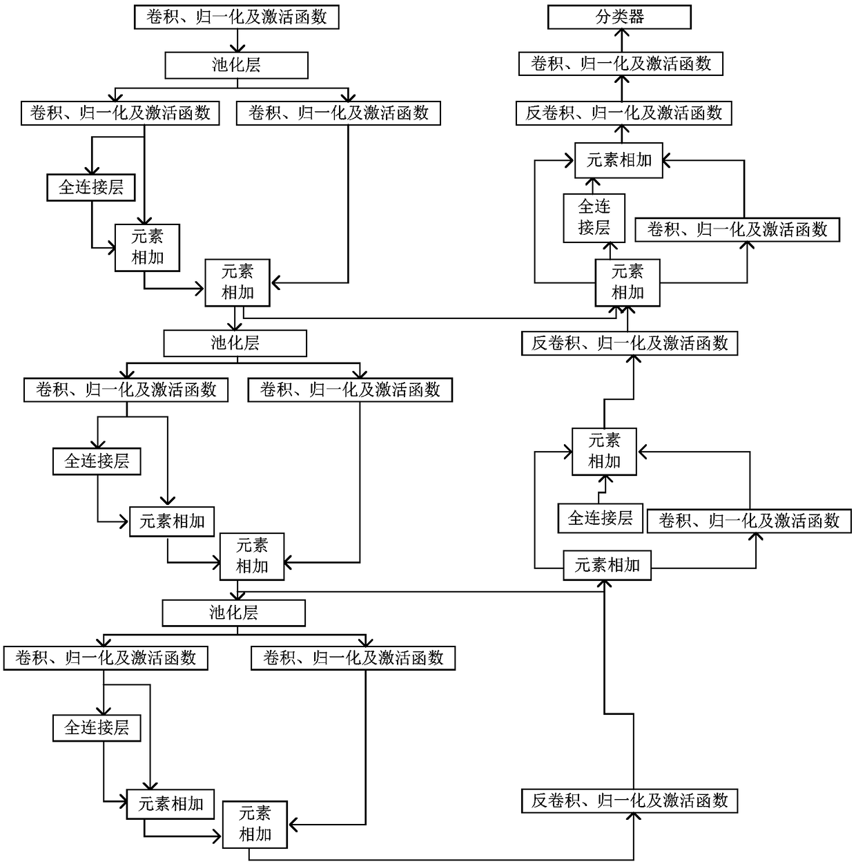
[0042] figure 1 For the network structure of the present invention, in the down-sampling stage, the original feature is continuously passed to each residual block to increase the reuse of the original feature, and it is passed to the up-sampling stage horizontally. In the downsampling stage, the present invention connects two convolutional layers and a feature map generated by a fully connected layer to form a network unit containing two parallel convolutions and a fully connected layer. This structure resamples the convolutional features extracted at a single scale, fuses multi-scale features, and incorporates global contextual information into the model. After convolution, a fully connected layer is used to avoid missing feature information during convolution. Since the image size of each downsampling stage is different, the expansion rate...


PUM


Abstract

The invention relates to a multi-scale nasopharyngeal tumor segmentation method based on CNN. Includes collecting MRI image data of nasopharyngeal region of several cases with nasopharyngeal tumor; Performing artificial edge labeling on the lesion area of the MRI image data collected in the previous step as label data layer by layer; Performing standardized preprocessing on the label data obtainedin the previous step and converting the label data into a two-dimensional data set; A CNN-based multi-layer two-dimensional convolution neural network is constructed and trained by using the two-dimensional data set in the previous step. For the MRI image data of nasopharyngeal region to be segmented, medical images of the same region and the same mode are collected, and the collected images arestandardized. The MRI image data of nasopharyngeal region to be segmented is segmented automatically by the network model. The invention can realize automatic segmentation of nasopharyngeal tumor, andcan obtain higher precision compared with mainstream network.

Description

technical field [0001] The present invention relates to the technical field of nasopharyngeal tumor image segmentation in the field of image segmentation, in particular to a CNN-based multi-scale nasopharyngeal tumor segmentation method. Background technique [0002] Nasopharyngeal carcinoma refers to malignant tumors that occur on the roof and side walls of the nasopharyngeal cavity. It is one of the high-incidence malignant tumors in my country, and its incidence rate is the first among ear, nose and throat malignant tumors. Nasopharyngeal tumors have a more complex anatomy than other types of tumors. Nasopharyngeal tumors are spatially similar to several tissues (air, bone, muscle, and mucous membrane) that process similar image intensities, and the shape and size of nasopharyngeal carcinomas, as well as non-uniform tumor intensities, are quite different. Therefore, it is necessary to design specific segmentation method. Moreover, the traditional diagnosis of nasophary...

Claims


Application Information

Patent Timeline
Patent Type & Authority Applications(China)
IPC IPC(8): G06T7/00G06T7/187
CPCG06T7/0012G06T2207/10088G06T2207/20081G06T2207/20084G06T2207/30096G06T7/187
Inventor 李孝杰罗超史沧红何嘉伍贤宇郭峰张宪刘书樵李俊良
Owner CHENGDU UNIV OF INFORMATION TECH
Why Eureka
Try Eureka