Visual surgical assembly and corresponding endoscope

An assembly and surgery technology, applied in the field of medical devices, can solve the problems of high resolution of visual images and no special requirements for the distance of light, so as to avoid cross infection and reduce costs

Pending Publication Date: 2019-03-22
SHANGHAI OUTAI MEDICAL EQUIP
View PDF0 Cites 4 Cited by
  • Summary
  • Abstract
  • Description
  • Claims
  • Application Information

AI Technical Summary

Problems solved by technology

[0005] Since the existing visual human flow endoscope can only meet the requirements of using the drainage tube to suck the embryo after observing the positioning of the embryo, so this kind of product has no special requirements for the high resolution of the sensor of the visual image and the distance of the light

Method used


Image

  • Visual surgical assembly and corresponding endoscope
  • Visual surgical assembly and corresponding endoscope
  • Visual surgical assembly and corresponding endoscope

Examples


Embodiment Construction

[0063] In order to describe the technical content of the present invention more clearly, further description will be given below in conjunction with specific embodiments.

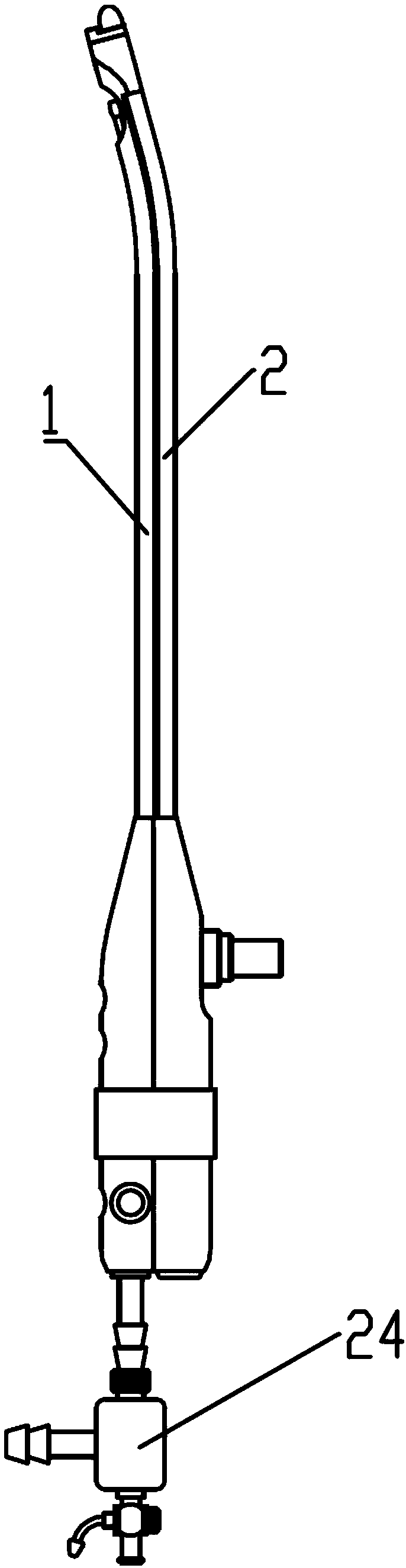
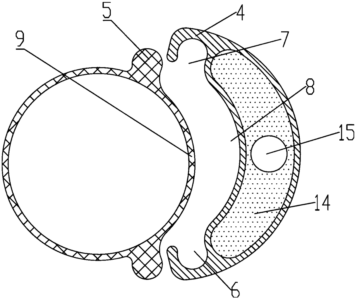
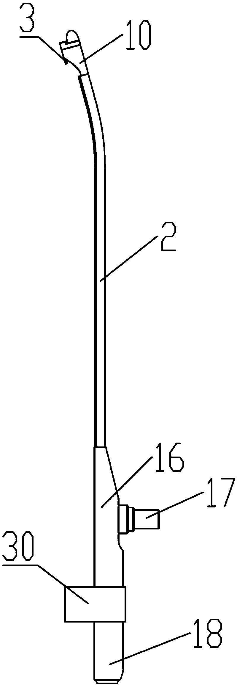
[0064] like Figure 1-8 As shown, it is an embodiment of a visualization surgery assembly provided by the present invention, which includes a disposable drainage tube 1 and a functional tube 2 for providing visualization functions. The disposable drainage tube and functional tube are along the tube body. Connected detachably in the axial direction, the functional tube is provided with a self-destruct part 3 for cutting the disposable drainage tube.

[0065] The assembly provided by the present invention is preferably used in artificial abortion surgery, and all components need to be sterilized before use. Among them, the functional tube can use steel structure, which can be reused after repeated disinfection. It can be used only after it meets the requirements of medical regulations and cannot be sterilize...


PUM


Abstract

The invention relates to a visual surgical assembly which comprises a disposable drainage tube and a functional tube. The functional tube is used for providing visual functions, the disposable drainage tube and the functional tube are detachably connected with each other along the axial direction of a tube, the functional tube is provided with a self-destruction component, and the self-destructioncomponent is used for breaking the disposable drainage tube by means of cutting. The invention further provides an endoscope. The visual surgical assembly and the endoscope have the advantages that the visual surgical assembly is of an assembled structure with the disposable drainage tube and the functional tube capable of providing the visual functions, the disposable drainage tube is detachablyconnected with the functional tube, the functional tube can be repeatedly disinfected and used, accordingly, the cost can be reduced, and risks of cross infection can be prevented.

Description

technical field [0001] The present invention relates to the technical field of medical devices, in particular to a visualized medical device, in particular to a visualized surgery assembly and a corresponding endoscope for artificial abortion. Background technique [0002] In the clinical operation of visualized abortion, it is often necessary to use a visualized endoscope for the observation, positioning and aspiration of early embryos. In the early abortion operations, due to the difficulty in realizing visualization, the metal tube with a channel was basically connected to the suction device at the rear end, and the surface mucosa of the uterine body was sucked out in the uterine cavity to achieve continued conception of the aborted embryo. Due to the inability to accurately locate the embryo in the uterine cavity, a total aspiration operation of the inner surface mucosa of the uterine cavity is generally performed, but the damage to the non-embryonic mucosa layer cannot ...

Claims


Application Information

Patent Timeline
Patent Type & Authority Applications(China)
IPC IPC(8): A61B17/42A61B90/00A61M1/00A61B1/05
CPCA61M1/0023A61B1/05A61B17/4241A61B90/37A61M2205/587A61M1/84A61M1/85A61M1/87A61B17/42A61B2017/4216A61B2017/0023A61B2017/00296A61B17/46A61B1/0014A61B1/015A61B1/00094A61B1/00062A61B1/07
Inventor 徐睿刘素萍汤竹君赵彤岑磊刘云贵王旭开成强
Owner SHANGHAI OUTAI MEDICAL EQUIP
Why Eureka
Try Eureka