A CT image pulmonary parenchyma three-dimensional semantic segmentation method based on a deep neural network

A deep neural network and CT image technology, applied in image segmentation, the field of three-dimensional semantic segmentation of CT image lung parenchyma based on deep neural network, can solve the problems of noise sensitivity, inability to make full use of the special information of volume data, and the effect is not optimal

Active Publication Date: 2019-04-09
BEIJING UNIV OF TECH
View PDF8 Cites 58 Cited by
  • Summary
  • Abstract
  • Description
  • Claims
  • Application Information

AI Technical Summary

Problems solved by technology

The shortcomings of the region growing method are: (1) how to define the regional consistency criterion; (2) the segmentation result has a great relationship with the selection of the seed point; (3) this method is also very sensitive to noise and may Formation of porous or even discontinuous regions at all
[0008] Although these 2D CNN-based methods gre

Method used


Image

  • A CT image pulmonary parenchyma three-dimensional semantic segmentation method based on a deep neural network
  • A CT image pulmonary parenchyma three-dimensional semantic segmentation method based on a deep neural network
  • A CT image pulmonary parenchyma three-dimensional semantic segmentation method based on a deep neural network

Examples


Embodiment Construction

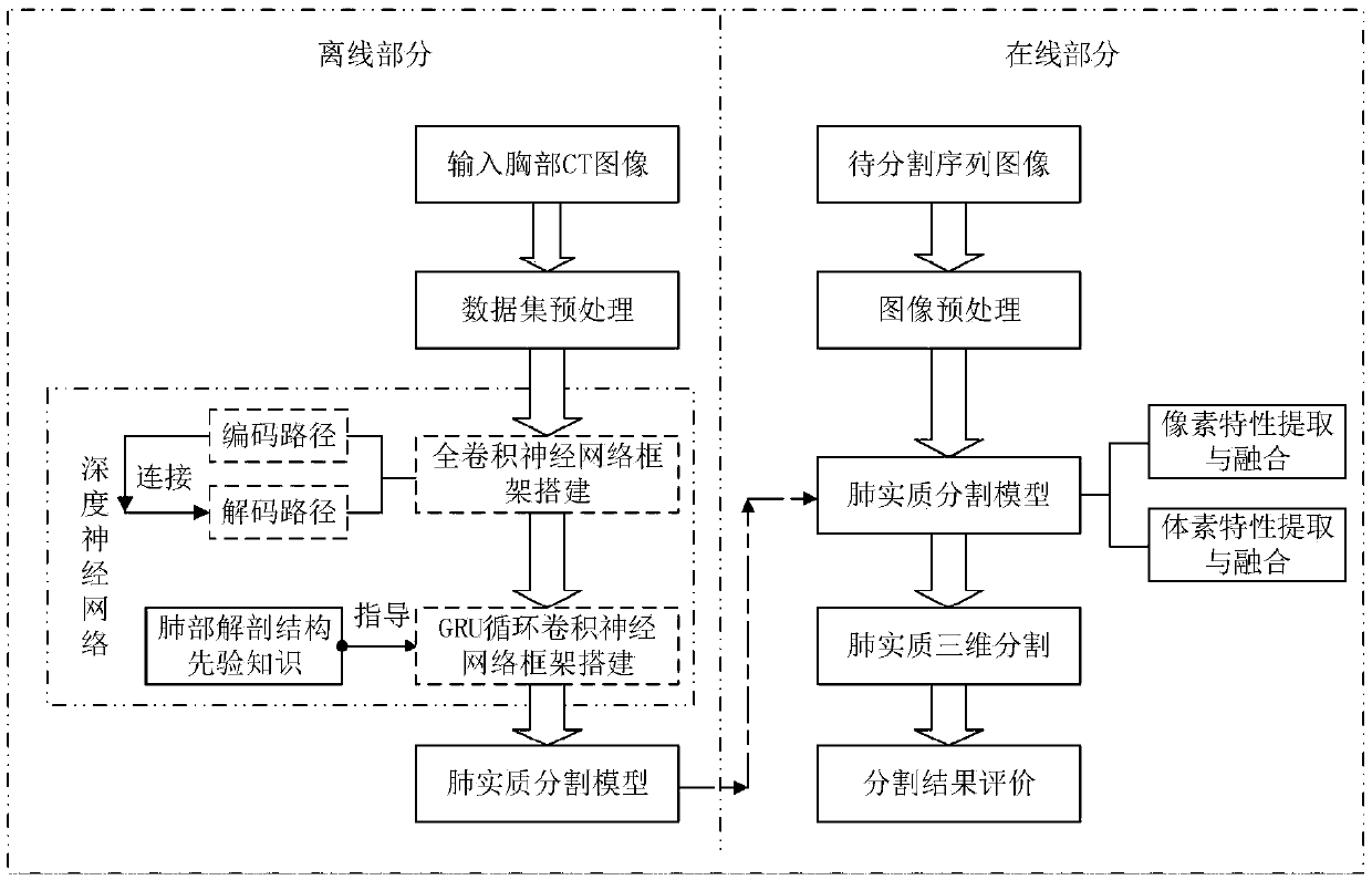
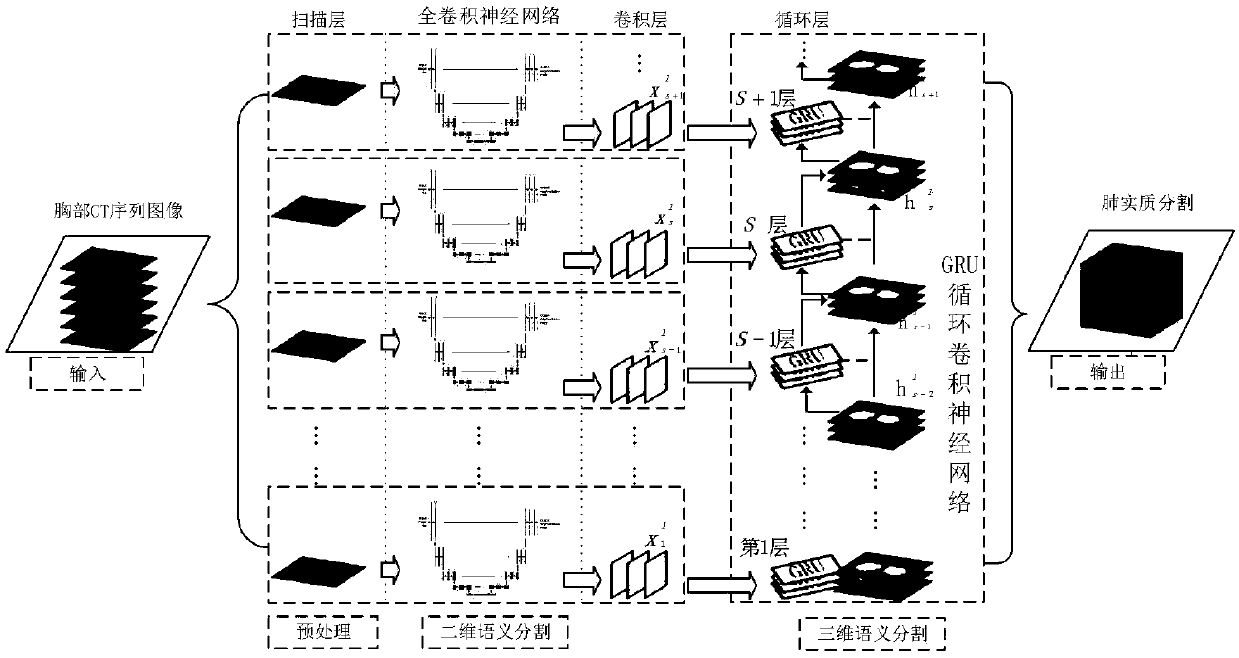
[0060]Below in conjunction with accompanying drawing of description, the embodiment of the present invention is described in detail:

[0061] The offline part is divided into 4 steps:

[0062] Step 1 Preprocessing of the dataset

[0063] The image I is normalized by the following formula using the window width and window level.

[0064]

[0065]

[0066]

[0067] In the formula, Winc is the WindowCenter read from the image, indicating the window level of the image; Winw is the WindowWidth read from the image, indicating the window width of the image; R is the RescaleSlope read from the image, indicating the pixel value RI is the RescaleIntercept read from the image, which represents the scale parameter of the pixel value; i represents each pixel of the image; N is the normalization function.

[0068] Then the images of different sizes are uniformly cropped to a size of 508×508 to form a unified sequence image. Finally, experienced doctors manually label the lung pa...


PUM


Abstract

The invention discloses a CT image pulmonary parenchyma three-dimensional semantic segmentation method based on a deep neural network. The segmentation method comprises an offline part and an online part. The offline part comprises four steps: preprocessing a data set; Constructing a full convolutional neural network framework; Constructing a GRU cyclic convolutional neural network framework; Network training. The online part comprises the following five steps: preprocessing an image; Extracting and fusing pixel features; Extracting and fusing voxel characteristics; Performing segmented output; Segmentation result evaluation. The feature fusion comprises feature fusion between an encoding layer and a decoding layer and feature fusion between anatomical structure layers. A deep neural network model with a gating circulation unit is designed, and spatial features are extracted by using anatomical structure prior information of a lung to effectively represent an apparent evolution relationship between fault sequences, so that accurate three-dimensional semantic segmentation is carried out on lung parenchyma in a CT image.

Description

technical field [0001] The invention belongs to the image segmentation technology in the field of medical image processing, in particular to a three-dimensional semantic segmentation method of lung parenchyma in CT images based on deep neural networks. Background technique [0002] Lung disease has always been a serious threat to human health. Lung cancer has the highest mortality rate of all malignant tumors, and the overall 5-year survival rate does not exceed 15%. With the rapid development of my country's economic level, people's attention to their own health is also increasing. Early diagnosis and early treatment of diseases have increasingly become the common concern of the whole society. In order to reduce the workload of doctors and detect lung diseases more quickly and accurately, the application of medical image processing technology in the auxiliary diagnosis of lung diseases is of great significance to the auxiliary diagnosis of clinical lung diseases. The segme...

Claims


Application Information

Patent Timeline
IPC IPC(8): G06T7/11G06K9/62G06N3/04G06N3/08
CPCG06N3/084G06T7/11G06T2207/10081G06T2207/20084G06T2207/20081G06T2207/30064G06N3/045G06F18/2414G06F18/253
Inventor 张辉张岩卓力李晓光
Owner BEIJING UNIV OF TECH
Why Eureka
Try Eureka