
Image processing method, image processing device and nuclear magnetic resonance imaging equipment

An image processing and image technology, applied in the interdisciplinary field of neuroscience and medical image processing, can solve the problems of difficult image fusion and different dimensions

Active Publication Date: 2019-07-16
TSINGHUA UNIV
View PDF4 Cites 6 Cited by
  • Summary
  • Abstract
  • Description
  • Claims
  • Application Information

AI Technical Summary

Problems solved by technology

But the dimensions of the three images are different: the anisotropic image is a two-dimensional image, the nerve distribution image is a three-dimensional image, and the diffusion envelope image is between two-dimensional and three-dimensional (the voxels to be measured are distributed on a two-dimensional plane, The diffusion envelope image in each voxel is three-dimensional), which causes some difficulties in image fusion

Method used


Image

  • Image processing method, image processing device and nuclear magnetic resonance imaging equipment
  • Image processing method, image processing device and nuclear magnetic resonance imaging equipment
  • Image processing method, image processing device and nuclear magnetic resonance imaging equipment

Examples


Embodiment Construction

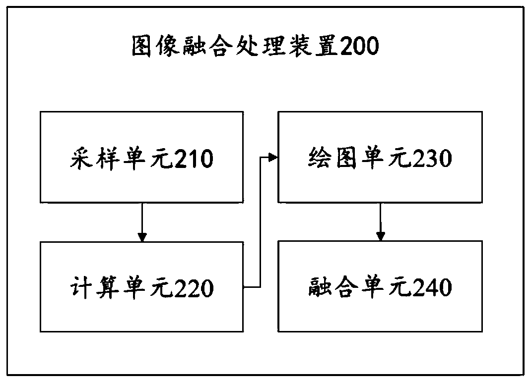
[0028] In order to make the object, technical solution and advantages of the present invention clearer, the present invention will be described in further detail below in conjunction with the accompanying drawings and embodiments. It should be understood that the specific embodiments described here are only used to explain the present invention, not to limit the present invention.

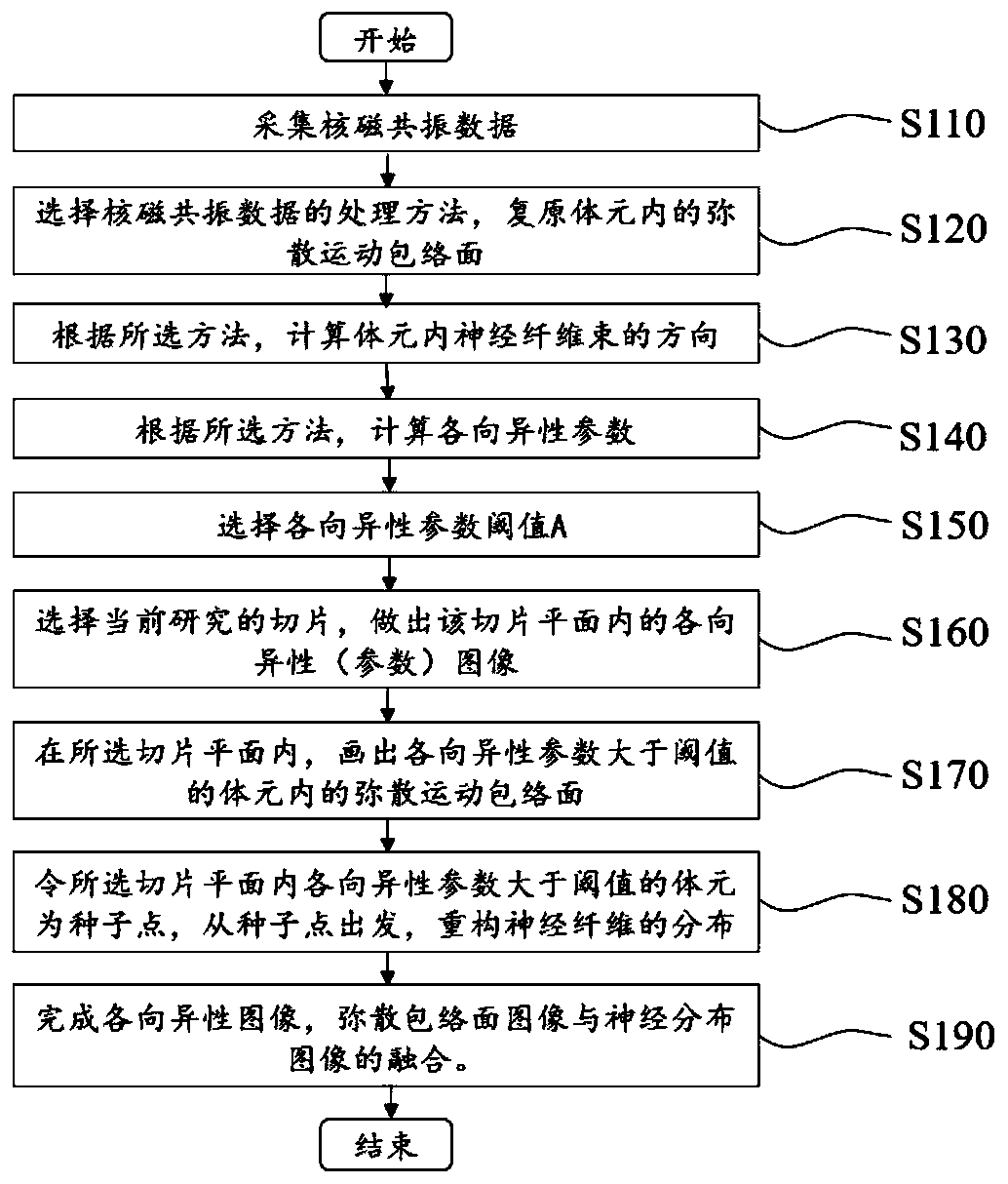
[0029] Such as figure 1 Shown is the flowchart of the image fusion method of the present invention, and it comprises the following steps:

[0030] Step S110: collecting the nuclear magnetic resonance data of the subject. Select a limited number of sampling directions on the unit sphere, and use nuclear magnetic resonance technology to measure the attenuation strength of the signal in the sampling direction in each voxel, that is, the nuclear magnetic resonance data.

[0031] Step S120: Select a processing method for the nuclear magnetic resonance data, and obtain an envelope of diffusion motion i...


PUM


Abstract

The invention relates to a medical imaging technology, in particular to a method for realizing fusion of an anisotropic image, a diffusion envelope surface image and a nerve distribution image. The method comprises the steps that corresponding anisotropic images, diffusion envelope surface images and nerve distribution images are acquired according to nuclear magnetic resonance data, and image fusion is completed by combining respective characteristics of the three images. The method is suitable for analyzing and reconstructing the complex biological tissue structure in the brain, and has important application values in the aspects of brain science, neuroscience, medical imaging and the like.

Description

technical field [0001] The present invention relates to the cross field of neuroscience and medical image processing, specifically, it relates to processing anisotropic images in nuclear magnetic resonance imaging, a method for fusing diffusion envelope images and nerve distribution images and a corresponding image processing device, and includes the Image processing device for magnetic resonance imaging equipment. The method and device are suitable for the research of brain structure and function, the diagnosis and treatment of brain diseases, and the preoperative planning of clinical neurosurgery operations, etc. Background technique [0002] Magnetic resonance imaging (MRI) uses the principle of nuclear magnetic resonance. According to the different attenuation of the released energy in different structural environments inside the material, the position of the atomic nucleus that constitutes the object can be known by detecting the emitted electromagnetic waves with an ex...

Claims


Application Information

Patent Timeline
Patent Type & Authority Applications(China)
IPC IPC(8): G06T5/50G01N24/08
CPCG01N24/08G06T5/50G06T2207/20221
Inventor 郑泉水史迪威
Owner TSINGHUA UNIV
Learn More