Ultrahigh-pixel tissue pathological image segmentation method

A technology for histopathology and image segmentation, applied in the field of ultra-high pixel histopathological image segmentation and ultra-high pixel tissue pathological image analysis, it can solve the problem of high computing time and cost, and it is difficult to obtain smooth and fine lesion area edges, influences, etc. problem, to achieve the effect of accurate and precise area segmentation

Active Publication Date: 2019-09-27
CHONGQING INST OF GREEN & INTELLIGENT TECH CHINESE ACADEMY OF SCI
View PDF7 Cites 8 Cited by
  • Summary
  • Abstract
  • Description
  • Claims
  • Application Information

AI Technical Summary

Problems solved by technology

[0003] At present, neural network deep learning technology has been widely used in this field. Although the existing methods based on image block segmentation have achieved certain results, however, for pathological slice images with ultra-high pixels, if smaller image blocks (such as 224 ×224 size) in the ultra-high pixel image for dense sampling method to detect the lesion area of ​​the whole slice, it often needs millions of dense sampling and repeated convolution operations, the calculation time and cost are relatively high, and in a certain To some extent, it affects the actual application effect; at the same time, the essence of the image block sampling method is to apply the image classification method to the task of segmenting the lesion area. Since it is difficult to achieve accurate area segmentation at the pixel level of pathological images, if the image When the sampling accuracy is not high, it is often difficult to obtain a smooth and fine edge of the lesion area

Method used


Image

  • Ultrahigh-pixel tissue pathological image segmentation method
  • Ultrahigh-pixel tissue pathological image segmentation method
  • Ultrahigh-pixel tissue pathological image segmentation method

Examples


Embodiment

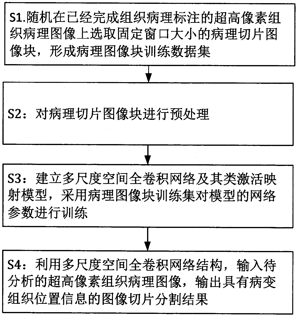
[0022] Multiple ultra-high pixel histopathological images taken clinically in the hospital, and histopathological annotations have been performed by professionals. For the newly captured ultra high pixel histopathological images, this embodiment provides an ultra high pixel histopathological image segmentation method , combined with figure 1 , the method consists of the following steps:

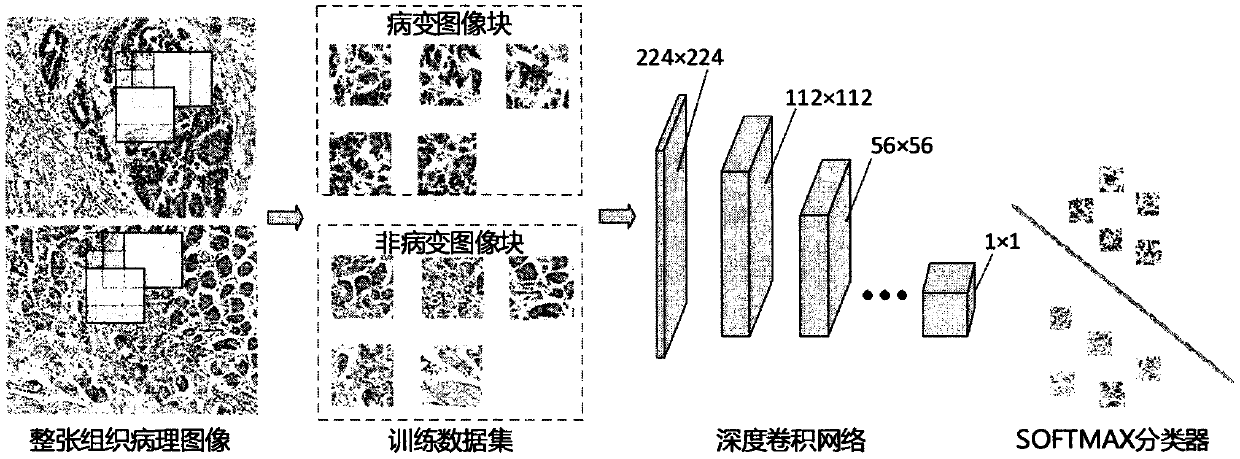
[0023] Step 1: Randomly select pathological slice image blocks with a fixed window size of 224×224 or 336×336 pixels on the ultra-high pixel histopathological images that have been histopathologically annotated at a step of 64 pixels to form a pathological image block training data set , where the data set is divided into tumor lesion and normal according to whether it contains diseased tissue.

[0024] Step 2: Delete the completely blank pathological slice image blocks from the training set, and at the same time, carry out conventional processing such as mean removal, normalization, princip...


PUM


Abstract

The invention discloses an ultrahigh-pixel tissue pathological image segmentation method, and belongs to the field of image processing and artificial intelligence. The method comprises the following steps of: S1, randomly selecting pathological section image blocks with fixed window sizes on an ultrahigh-pixel tissue pathological image on which tissue pathological labeling is completed to form a pathological image block training data set; S2, preprocessing the pathological section image blocks; s3, establishing a multi-scale space full convolution network and a class activation mapping model thereof, and training network parameters of the model by adopting a pathological image block training set in combination with a full connection layer so as to realize accurate lesion classification and recognition based on image blocks; and S4, inputting the to-be-analyzed ultra-high pixel tissue pathological image by using the multi-scale space full convolution network structure, and outputting an image slice segmentation result with pathological tissue position information. The ultra-high pixel tissue pathological image segmentation method can efficiently and accurately realize pixel-level accurate region segmentation of the ultra-high pixel tissue pathological image.

Description

technical field [0001] The invention relates to an ultra-high pixel histopathological image segmentation method, which belongs to the field of image processing and artificial intelligence, and is especially suitable for ultra-high pixel histopathological image analysis. Background technique [0002] Histopathological evaluation is essential for cancer diagnosis. By observing the tissue slice images of actual patients, pathologists can accurately judge the patient's condition. Computer vision-based automatic diagnosis of histopathology helps to reduce the workload of pathologists. In recent years, researchers working in this field have made remarkable achievements. The size of ultra-high-pixel histopathological images often exceeds 100 million pixels (usually greater than 100,000×100,000 pixels), and a patient often needs to collect multiple full-section images to determine his condition. Perception often requires millions of dense sampling and repeated convolution operati...

Claims


Application Information

Patent Timeline
Patent Type & Authority Applications(China)
IPC IPC(8): G06T7/11G06N3/04
CPCG06T7/11G06T2207/20081G06T2207/20084G06T2207/30004G06N3/045
Inventor 陈琳彭彬彬尚明生朱帆
Owner CHONGQING INST OF GREEN & INTELLIGENT TECH CHINESE ACADEMY OF SCI
Why Eureka
Try Eureka