Supercharge Your Innovation With Domain-Expert AI Agents!

Fixing support for radiography in dentistry and method

A technology for fixing brackets and film brackets, which is applied in the fields of radiological diagnosis instruments, applications, medical science, etc., can solve the problems of large structure of equipment, insufficient film strength or trembling, blurring, etc., to achieve the effect of fixing and strengthening

Pending Publication Date: 2019-10-11
上海东方肝胆外科医院
View PDF0 Cites 0 Cited by
  • Summary
  • Abstract
  • Description
  • Claims
  • Application Information

AI Technical Summary

Problems solved by technology

On the one hand, this way of filming is prone to unclear shooting due to the position of the finger, or the problem of poor filming effect due to the movement of the film when it is held by the finger
On the other hand, due to children or the elderly, inadequacies or tremors occur in the process of resisting the film, resulting in offset or blurred photographs
[0003] In addition, in the existing technology, in order to avoid the deviation of the imaging angle, a spherical imaging bracket is adopted. The imaging bracket has a connecting rod, and one end of the connecting rod extends into the oral cavity to reach the The position of the tooth end, and the other end is connected to a ring. Although this structure makes the center of the film and the ring on the same line to facilitate shooting, but because this type of equipment is large and complex in structure, it must be connected to the oral cavity when it is used. , so that the patient has to make some uncomfortable postures in order to match the position of the spherical bracket. Moreover, when the mouth is forcibly fixed, nausea and retching often occur, which is not conducive to the normal progress of radiography. And the experience is very bad, and the inside of the mouth is easily scratched by this kind of equipment

Method used


Image

  • Fixing support for radiography in dentistry and method
  • Fixing support for radiography in dentistry and method
  • Fixing support for radiography in dentistry and method

Examples


Embodiment 1

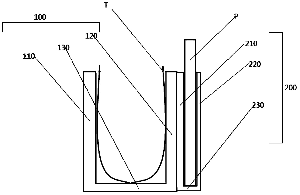
[0075] Such as image 3 shown, for figure 1 A schematic diagram of the cross-sectional structure along the direction of A-A.

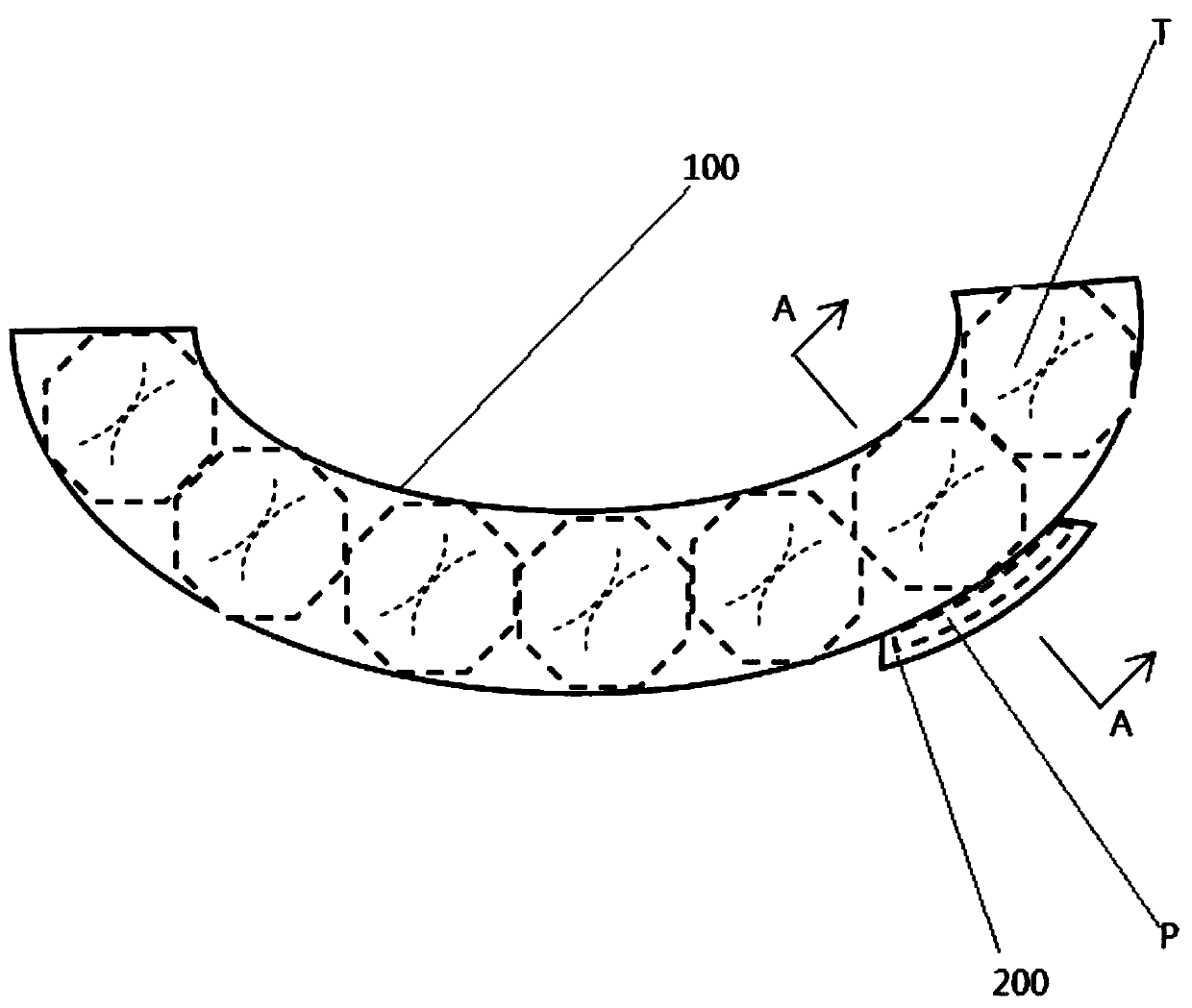
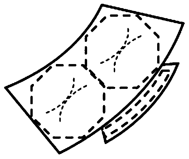
[0076] In this embodiment, the tooth bracket 100 is composed of the inner side plate 120 of the tooth bracket, the outer side plate 110 of the tooth bracket and the groove bottom plate 130 of the tooth bracket;

[0077] The inner side plate 120 of the tooth bracket 100 and the outer side plate 110 of the tooth bracket are installed on both sides of the bottom plate 130 of the tooth bracket groove, forming a U-shaped groove I, and the tooth T is clamped therein during use;

[0078] Considering the size difference of human teeth, the device can be made of deformable materials such as silicon rubber in addition to being made into various models. The brace fits better around the tooth so it doesn't fall out during the photoshoot.

[0079] The device can also be made of elastic materials, generally using a bracket slightly smaller than the teeth. When it...

Embodiment 2

[0089] Such as Figure 8 shown, for figure 1 A schematic diagram of the cross-sectional structure along the direction of A-A.

[0090] In this embodiment, the tooth bracket 100 is composed of the inner side plate 120 of the tooth bracket, the outer side plate 110 of the tooth bracket and the groove bottom plate 130 of the tooth bracket;

[0091] The inner side plate 120 of the tooth bracket 100 and the outer side plate 110 of the tooth bracket are installed on both sides of the bottom plate 130 of the tooth bracket groove, forming a U-shaped groove I, and the tooth T is clamped therein during use;

[0092] Considering the size difference of human teeth, the device can be made of deformable materials such as silicon rubber in addition to being made into various models. The brace fits better around the tooth so it doesn't fall out during the photoshoot.

[0093] The device can also be made of elastic materials, generally using a bracket slightly smaller than the teeth. When i...

Embodiment 3

[0106] Such as Figure 4 shown, for figure 1 A schematic diagram of the cross-sectional structure along the direction of A-A.

[0107] In this embodiment, the tooth bracket is composed of the inner side plate 120 of the tooth bracket, the outer side plate 110 of the tooth bracket and the groove bottom plate 130 of the tooth bracket;

[0108] The inner side plate 120 of the tooth bracket and the outer side plate 110 of the tooth bracket are installed on both sides of the bottom plate 130 of the tooth bracket groove, forming a U-shaped groove I, and the tooth T is clamped therein during use;

[0109] Considering the difference in the size of human teeth, the device can be made of deformable materials such as silicon rubber in addition to being made into various models. When it is set outside the teeth, it can make the teeth The brace fits better around the tooth so it doesn't fall out during the photoshoot.

[0110] The device can also be made of elastic materials, generally ...


PUM

PropertyMeasurementUnit
Thicknessaaaaaaaaaa

Abstract

The invention provides a fixing support for radiography in the dentistry. The fixing support is installed on the teeth and used for containing a film during radiography of the teeth. The fixing support is characterized by comprising a tooth support body and a radiography support body; the tooth support body is composed of a tooth support body inner side plate, a tooth support body outer side plateand a tooth support body groove baseplate; the tooth support body inner side plate and the tooth support body outer side plate are installed at the two sides of the tooth support body groove baseplate to form a U-shaped groove I; the radiography support body is composed of a radiography support body inner side plate, a radiography support body outer side plate and a radiography support body groove baseplate; the radiography support body inner side plate and the radiography support body outer side plate are installed at the two sides of the radiography support body groove baseplate to form a U-shaped groove II; the radiography support body inner side plate and the tooth support body outer side plate are installed in an integrated two-face mode or are detachably or fixedly installed; the U-shaped groove I is parallel to the U-shaped groove II. The fixing support for radiography in the dentistry can be easily installed on the teeth of the human body, and the radiography effect can be achieved without hand holding or external force abutting.

Description

technical field [0001] The invention relates to the field of auxiliary equipment for dental radiographs, in particular to a fixed bracket for dental radiographs and a simple radiographing method using the bracket. Background technique [0002] In the existing medical process, when it is necessary to take a film test on the teeth, the patient often needs to hold the film in the mouth with his hands to carry out. On the one hand, this way of filming is prone to unclear shooting due to finger position problems, or poor filming effect due to movement of the film during the process of being held against by fingers. On the other hand, due to children or old people, the problem of insufficient force or trembling occurs in the process of resisting the film, resulting in offset or blurred photographs. [0003] In addition, in the existing technology, in order to avoid the deviation of the imaging angle, a spherical imaging bracket is adopted. The imaging bracket has a connecting rod...

Claims


Application Information

Patent Timeline
IPC IPC(8): A61B6/14
CPCA61B6/512
Inventor 高昊辰吴鉴今刘国勤周嘉骏郑星吕婕李琼杨萍孟子丰张凇睿
Owner 上海东方肝胆外科医院
Features
  • R&D
  • Intellectual Property
  • Life Sciences
  • Materials
  • Tech Scout
  • Unparalleled Data Quality
  • Higher Quality Content
  • 60% Fewer Hallucinations
Learn More