
A Semi-Automatic Brain Region Segmentation Method for 3D Cytoarchitectural Images

A three-dimensional cell and semi-automatic technology, applied in the field of image processing, to achieve the effect of lowering the threshold and improving efficiency

Active Publication Date: 2022-06-03
HUST SUZHOU INST FOR BRAINMATICS
View PDF5 Cites 0 Cited by
  • Summary
  • Abstract
  • Description
  • Claims
  • Application Information

AI Technical Summary

Problems solved by technology

For the extraction of such area boundaries, traditional image segmentation algorithms that rely on continuous and regular grayscale information are helpless

Method used


Image

  • A Semi-Automatic Brain Region Segmentation Method for 3D Cytoarchitectural Images
  • A Semi-Automatic Brain Region Segmentation Method for 3D Cytoarchitectural Images

Examples


Embodiment Construction

[0028] The technical solutions in the embodiments of the present invention will be clearly and completely described below with reference to the accompanying drawings in the embodiments of the present invention. Obviously, the described embodiments are only a part of the embodiments of the present invention, rather than all the embodiments. Based on the embodiments of the present invention, all other embodiments obtained by those of ordinary skill in the art without creative efforts shall fall within the protection scope of the present invention.

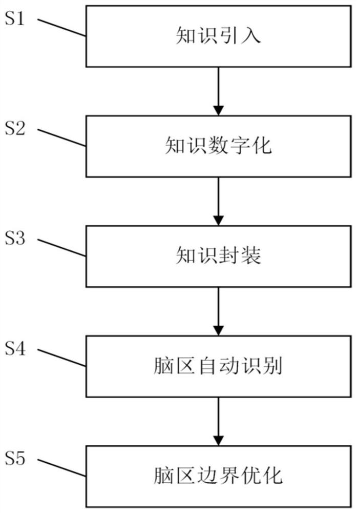
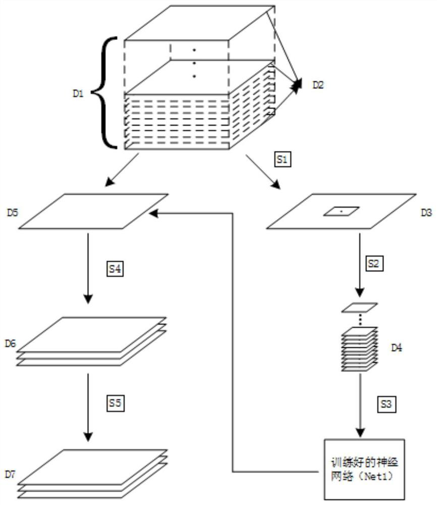
[0029] See figure 1 As shown, one embodiment of the present invention is a semi-automatic brain region segmentation method for three-dimensional cytoarchitecture images, and the method includes the following steps:

[0030] Step S1, knowledge introduction: constructing an image data set based on three-dimensional cells, selecting a two-dimensional image sequence, and marking the boundaries of each brain region on each image of the tw...


PUM


Abstract

The present invention provides a semi-automatic brain region segmentation method for three-dimensional cell-constructed images, comprising: step S1, knowledge introduction; step S2, knowledge digitization; step S3, knowledge encapsulation; step S4, automatic recognition of brain regions; step S5, brain region boundaries optimization. The present invention can better extract higher-dimensional and more abstract features through the deep learning method, so it can identify the overall boundary of the brain region composed of discrete cell bodies; in addition, through interactive segmentation, the present invention successfully integrates neuroanatomical The prior knowledge of the expert is introduced into the predictive network constructed by the deep learning technology. By digitizing the prior knowledge of neuroanatomy mastered by the expert, the experience that can only be understood in the brain of the expert can be turned into a tool and reused. It greatly reduces the threshold for automatic identification of brain regions, allowing ordinary neuroscience researchers to get rid of the high reliance on knowledge in the narrow field of neuroanatomy, and greatly improving the efficiency of brain region identification.

Description

technical field [0001] The invention relates to the field of image processing, in particular to a semi-automatic brain region segmentation method of a three-dimensional cytoarchitecture image with a resolution of 100 microns to microns. Background technique [0002] Brain imaging is one of the necessary technical means to carry out neuroscience research. With the advancement of microscopic optical imaging technology, a large amount of 3D brain image data has been acquired at the micron-resolution level, laying the foundation for research on a finer spatial scale. Among the many types of brain images, cytoarchitecture images are considered the gold standard for identifying different brain regions in the brain. In this type of image, the cell body morphology and spatial aggregation patterns of cells in different brain regions are different visible to the human eye. Therefore, experienced neuroanatomical experts usually find and manually draw each brain by carefully identifyin...

Claims


Application Information

Patent Timeline
Patent Type & Authority Patents(China)
IPC IPC(8): G06T7/12
CPCG06T7/12G06T2207/10056G06T2207/20081G06T2207/30016
Inventor 丰钊李安安刘鑫倪鸿龚辉骆清铭
Owner HUST SUZHOU INST FOR BRAINMATICS
Learn More