Transmembrane sensor to evaluate neuromuscular function

A sensor, sensor housing technology, applied in the direction of sensors, applications, surgical robots, etc., that can solve problems that do not allow reliable characterization of insertion activity, spontaneous activity, size and shape of motor units and/or interference patterns, large surface area, etc.

Pending Publication Date: 2020-04-10
POWELL MANSFIELD
View PDF10 Cites 2 Cited by
  • Summary
  • Abstract
  • Description
  • Claims
  • Application Information

AI Technical Summary

Problems solved by technology

However, due to the large surface area of ​​SEMG sensors, SEMG data may have limited spatial resolution relative to NEMG data
For example, SEMG data can be susceptible to mechanical and electrical artifacts and cr

Method used


Image

  • Transmembrane sensor to evaluate neuromuscular function
  • Transmembrane sensor to evaluate neuromuscular function
  • Transmembrane sensor to evaluate neuromuscular function

Examples


Embodiment Construction

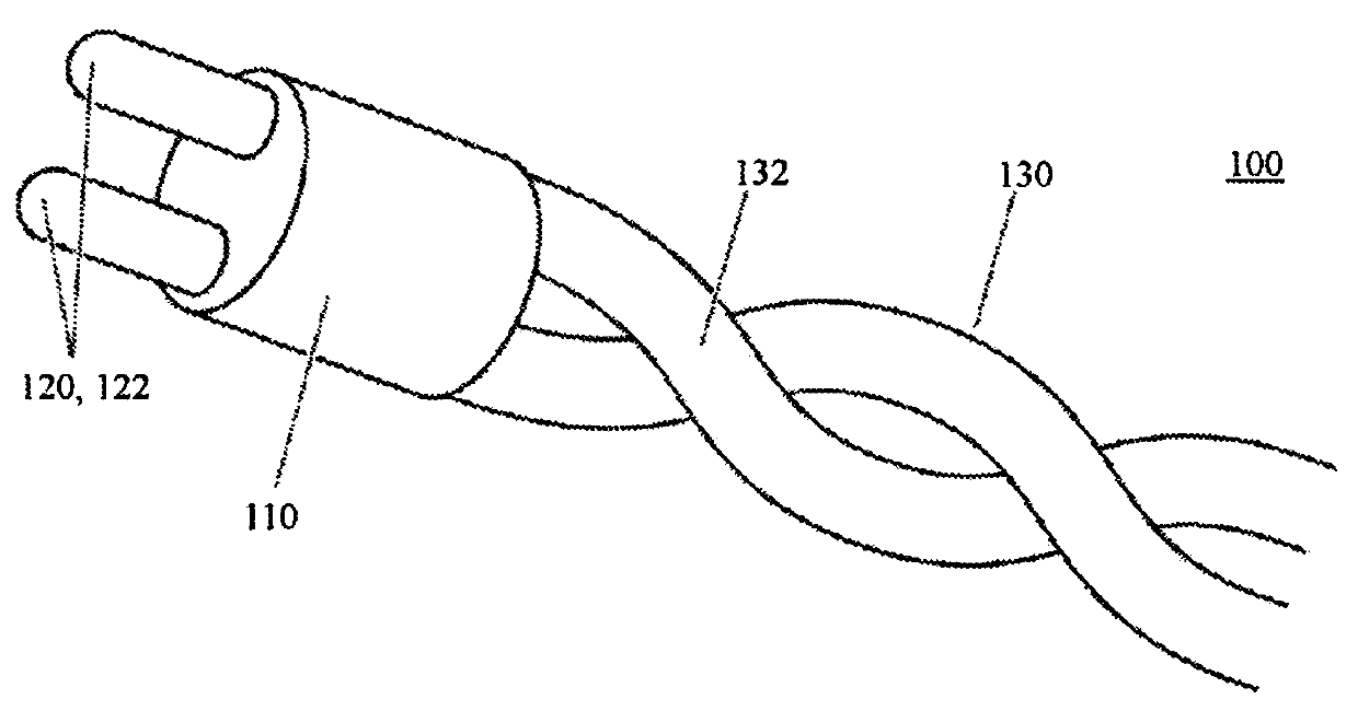
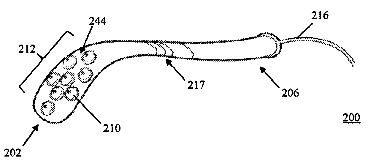
[0031] Described herein are sensor devices, systems, and methods for use in non-invasive diagnostic procedures for neuromuscular function of tissue within a body cavity or on the surface of an anatomical structure. In some variations, the sensor assembly can be used to measure electrical activity of one or more muscles. Generally, non-invasive transmembrane EMG (TM-EMG) sensors can be used to receive electrical activity signal data corresponding to a particular muscle, which signal data is used to generate EMG data. One or more sensors may be incorporated into one or more sensor arrays in the probe. The probe and sensor array can be configured to contact muscle tissue in membranous body cavities (eg, oropharynx, abdominal cavity, pelvic cavity, joint cavities) or other anatomical structures (eg, eyes), including anatomical structures accessed intraoperatively.

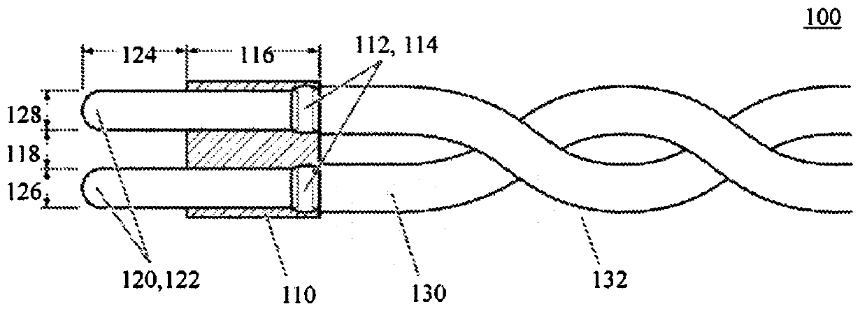
[0032] A sensor assembly as described herein may include one or more pairs of closely spaced atraumatic electrodes ...


PUM

PropertyMeasurementUnit
Diameteraaaaaaaaaa

Abstract

Devices, systems, and methods herein relate to electromyography (EMG) that may be used in diagnostic and/or therapeutic applications, including but not limited to electrophysiological study of musclesin the body relating to neuromuscular function and/or disorders. Sensor assemblies and methods are described herein for non-invasively generating an EMG signal corresponding to muscle tissue where the sensor may be positioned directly on a surface of the muscle tissue including any associated membrane (e.g., mucosal, endothelial, synovial) overlying the muscle tissue. A sensor assembly may include one or more pairs of closely spaced, atraumatic electrodes in a bipolar or multipolar configuration. The first and second electrodes may be applied against a surface of muscle tissue (that may include a membrane overlying the muscle) and receive electrical activity signal data corresponding to an electrical potential difference of the portion of muscle between the electrodes.

Description

[0001] Related applications [0002] This application claims the benefit of US Provisional Application 62 / 515,364, filed June 5, 2017, entitled "TRANSMEMBRANE SENSOR TO EVALUATE NEUROMUSCULAR FUNCTION," which is incorporated herein by reference in its entirety. technical field [0003] The devices, systems and methods herein relate to electromyography (EMG), which can be used for diagnostic and / or therapeutic applications including, but not limited to, electrophysiological studies of in vivo muscles related to neuromuscular function and / or disorders. Background technique [0004] EMG is related to the study of electrical activity that occurs in peripheral nerve and muscle tissue. There are generally two techniques for recording EMG signals: intramuscular or needle EMG (NEMG) and surface EMG (SEMG). Needle EMG surgery involves inserting needle electrodes directly into the muscle to be examined. Needle EMG is considered the clinical gold standard for assessing a range of neu...

Claims


Application Information

Patent Timeline
IPC IPC(8): A61B5/0488A61B5/04A61B5/00A61N1/04A61N1/05A61B5/0492A61B5/296
CPCA61B34/30A61B2562/046A61B5/30A61B5/24A61B5/296A61B5/389A61B5/6852A61B2562/16A61H2230/605A61B5/394
Inventor P·T·曼斯菲尔德C·D·斯图特T·J·科斯克
Owner POWELL MANSFIELD
Why Eureka
Try Eureka