Methods and devices for detecting biomarkers associated with preeclampsia
A biomarker and pre-eclampsia technology, applied in biochemical equipment and methods, measurement devices, microbial determination/testing, etc., can solve the problem of intensified identification of predictive biomarkers, comprehensive understanding of the pathogenesis of PE is out of reach, Difficulties in developing targeted therapy strategies
- Summary
- Abstract
- Description
- Claims
- Application Information
AI Technical Summary
Problems solved by technology
Method used
Image

Examples
Embodiment 1
[0314] Example 1: Human endometrial stromal cells from women with previous sPE pregnancies fail to decidualize in vitro
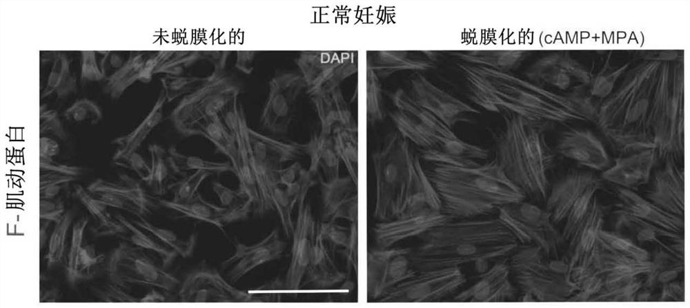
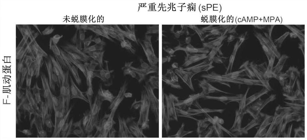
[0315] Decidualization of hESCs isolated from endometrial biopsy samples from patients with sPE in previous pregnancies (n=13) was assessed and compared to control patients with normal delivery outcomes (n=13). Table 2 summarizes the maternal and infant characteristics of the participants. hESCs were decidualized by treatment with cAMP and medroxyprogesterone acetate (MPA) for 5 days. As experimental controls, cells from the same donor were cultured in parallel in the absence of cAMP and MPA.
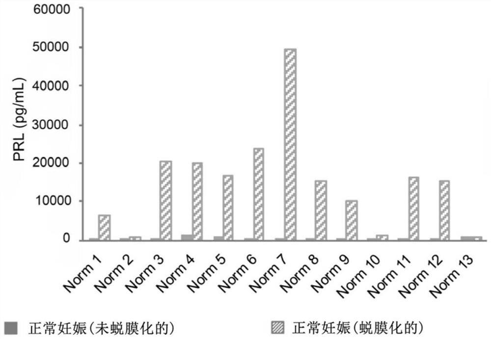
[0316] Localization of F-actin in decidual cells from non-abnormal pregnant women showed expected cytoskeletal remodeling and shape changes consistent with conversion of fibroblasts to the decidual phenotype ( Figure 1A ). In contrast, hESCs from women with sPE did not undergo these changes ( Figure 1B ). In non-decidualized hESCs, PRL detected in conditioned ...
Embodiment 2
[0318] Example 2: Alterations in the global transcriptional profile of decidualized hESCs from prior sPE patients.
[0319] To identify molecular changes in functional decidualization defects found in hESCs from women who had undergone sPE, a microarray strategy was used. Specifically, the transcriptomic analysis of non-decidualized and decidualized hESCs established from the normal pregnancy group and the sPE pregnancy group was performed in vitro ( Figure 2A ). Table 3 shows the clinical characteristics of the endometrial donors.
[0320] Figure 2B An overview of the results is given in . In the non-decidualized state, only 5 genes were differentially expressed between the control and sPE samples, and the fold difference was small ( Figure 2C ). Thus, at baseline, hESCs from patients with prior sPE were very similar to hESCs from control women.
[0321] The expression of 74 genes was significantly regulated ≥2-fold during decidualization in samples from control dono...
Embodiment 3
[0325] Example 3: In situ molecular defects in decidua basalis or parietal decidua from control versus sPE pregnancies
[0326] Isolate sections of the basal decidua (DB) or parietal decidua (DP) using laser microdissection. From gestational age-matched samples from women with sPE case versus control (from women with preterm birth (nPTB) without evidence of infection; Figure 3A ) to capture cells in tissue sections from biopsy samples. Table 8 summarizes the clinical characteristics of the participants.
[0327] Table 8. Maternal and Fetal Characteristics of Decidual Donors (Transcriptome Analysis of In Situ Expression of Decidual Genes in Severe Preeclampsia (sPE) vs. Spontaneous Preterm Birth Without Signs of Infection (NPTB))
[0328]
[0329] Mean ± SEM ** One-tailed Student's t-test
[0330] NA: not applicable
[0331] Figure 3B An overview of the results is shown in . In decidua basalis, 79 genes were significantly DE in sPE vs. nPTB, with smaller fold changes...
PUM
| Property | Measurement | Unit |
|---|---|---|
| transmittivity | aaaaa | aaaaa |
| transmittivity | aaaaa | aaaaa |
| transmittivity | aaaaa | aaaaa |
Abstract
Description
Claims
Application Information
 - R&D Engineer
- R&D Manager
- IP Professional
- Industry Leading Data Capabilities
- Powerful AI technology
- Patent DNA Extraction
Browse by: Latest US Patents, China's latest patents, Technical Efficacy Thesaurus, Application Domain, Technology Topic.
© 2024 PatSnap. All rights reserved.Legal|Privacy policy|Modern Slavery Act Transparency Statement|Sitemap