Hip joint CT image segmentation method and device, storage medium and computer equipment

A CT image and storage medium technology, applied in the computer field, can solve the problems of slow segmentation speed and low segmentation accuracy of CT images of hip joints, and achieve the effect of improving segmentation speed and segmentation accuracy

Pending Publication Date: 2020-09-29
BEIJING TINAVI MEDICAL TECH
View PDF7 Cites 5 Cited by
  • Summary
  • Abstract
  • Description
  • Claims
  • Application Information

AI Technical Summary

Problems solved by technology

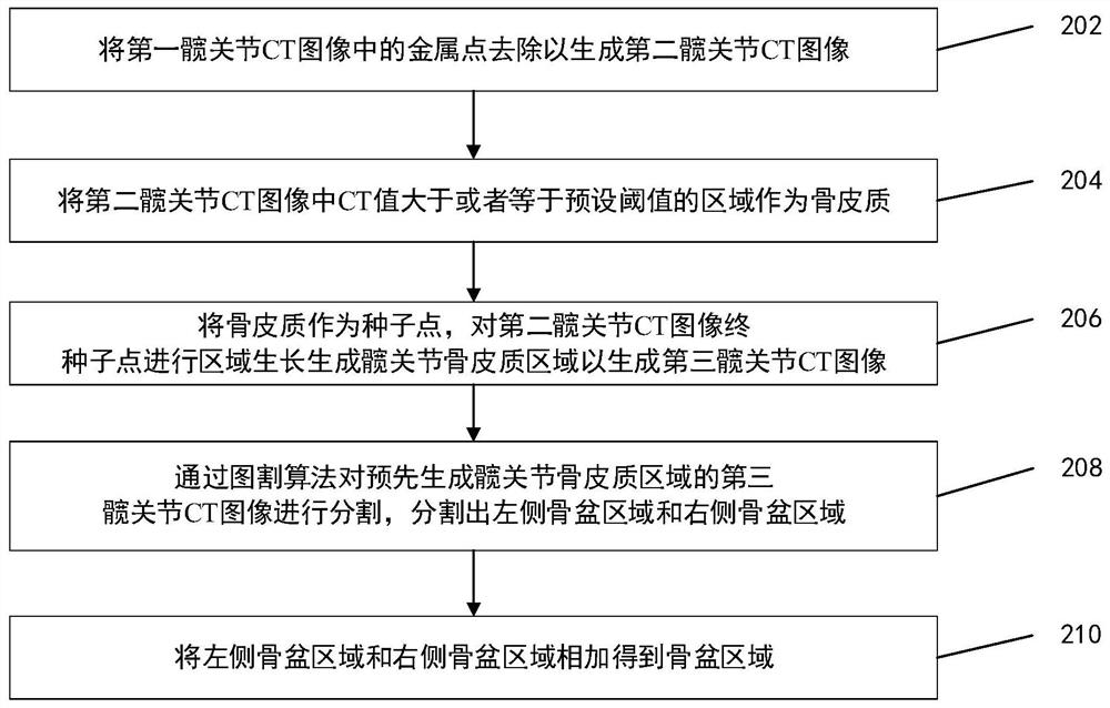
[0005] In view of this, the embodiments of the present invention provide a hip joint CT image segmentation method, device, storage medium and computer equipment, which can solve the problems of slow hip joint CT image segmentation and low segmentation accuracy in the related art

Method used


Image

  • Hip joint CT image segmentation method and device, storage medium and computer equipment
  • Hip joint CT image segmentation method and device, storage medium and computer equipment
  • Hip joint CT image segmentation method and device, storage medium and computer equipment

Examples


Embodiment Construction

[0035] In order to better understand the technical solutions of the present invention, the embodiments of the present invention will be described in detail below in conjunction with the accompanying drawings.

[0036] It should be clear that the described embodiments are only some of the embodiments of the present invention, not all of them. Based on the embodiments of the present invention, all other embodiments obtained by persons of ordinary skill in the art without creative efforts fall within the protection scope of the present invention.

[0037] Terms used in the embodiments of the present invention are only for the purpose of describing specific embodiments, and are not intended to limit the present invention. As used in the embodiments of the present invention and the appended claims, the singular forms "a", "said" and "the" are also intended to include the plural forms unless the context clearly indicates otherwise.

[0038] It should be understood that the term "an...


PUM


Abstract

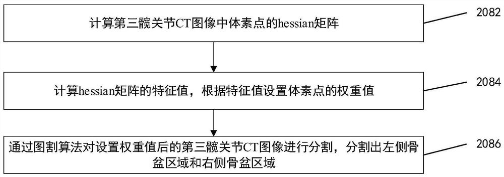
The embodiment of the invention provides a hip joint CT image segmentation method and device, a storage medium and computer equipment. The method provided by the embodiment of the invention comprisesthe steps of segmenting a third hip joint CT image of a hip joint bone cortex area generated in advance through a graph cutting algorithm, and a left side pelvis area, a right side pelvis area, a leftfemur area and a right femur area are segmented; and adding the left-side pelvis area and the right-side pelvis area to obtain a pelvis area. According to the method of the invention, the image segmentation algorithm is improved in the hip joint CT image segmentation process, and the segmentation speed and segmentation accuracy of the hip joint CT image can be improved.

Description

【Technical field】 [0001] The invention relates to the field of computers, in particular to a hip joint CT image segmentation method, device, storage medium and computer equipment. 【Background technique】 [0002] Image segmentation is an important part of medical image processing. In medicine, image segmentation is an essential step for subsequent operations such as feature extraction, quantitative analysis, and 3D reconstruction of normal or diseased tissue. In addition, many image-guided surgeries also require image segmentation as a prerequisite. In hip replacement surgery, using images to accurately segment the pelvic region, left femoral region, and right femoral region plays a vital role in registration and surgical planning in hip replacement surgery. [0003] The results of computed tomography (CT) imaging technology exist in the form of sequences. Therefore, CT images of hip joints usually contain hundreds of medical images, and the amount of data is large. If ma...

Claims


Application Information

Patent Timeline
Patent Type & Authority Applications(China)
IPC IPC(8): G06T7/11G06T7/136
CPCG06T7/11G06T7/136G06T2207/10081G06T2207/30008
Inventor 李亚赵永强
Owner BEIJING TINAVI MEDICAL TECH
Why Eureka
Try Eureka