Multimodal collaborative image segmentation system for esophageal cancer lesions based on self-sampling similarity
A multi-modal, esophageal cancer technology, applied in the field of medical image intelligent processing, can solve the problems that researchers only consider, and achieve the effect of improving efficiency and precise segmentation
- Summary
- Abstract
- Description
- Claims
- Application Information
AI Technical Summary
Problems solved by technology
Method used
Image
Examples
Embodiment Construction
[0034] The embodiments of the present invention will be described in detail below, but the protection scope of the present invention is not limited to the examples.
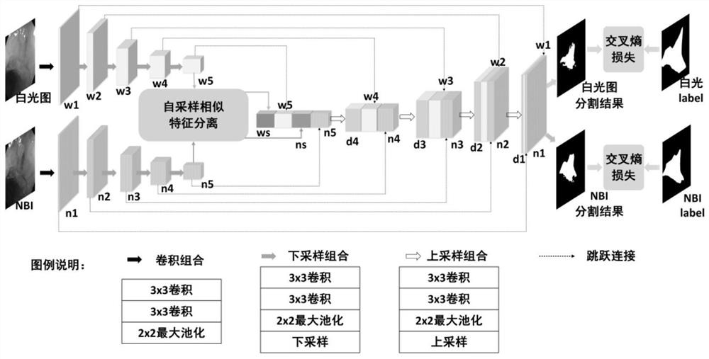
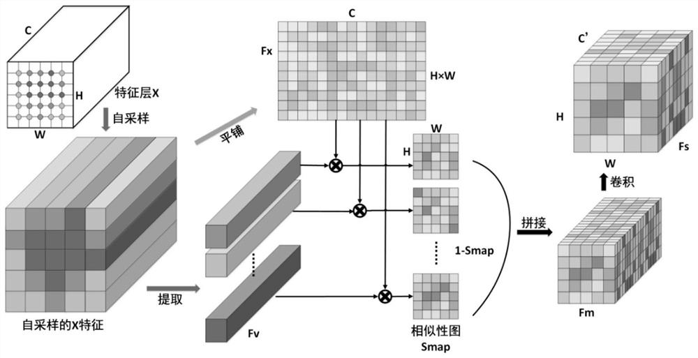
[0035] use figure 1 With the network structure in , a multimodal neural network is trained with 268 white-light NBI image pairs to obtain an automatic lesion region segmentation model.
[0036] The specific steps are:
[0037] (1) When training, scale the image to 500×500. Set the initial learning rate to 0.0001, the decay rate to 0.9, and decay once every two cycles. Minimize the loss function using mini-batch stochastic gradient descent. The batch size is set to 4. Update all parameters in the network, minimize the loss function of the network, and train until convergence;
[0038] (2) When testing, the image I Adjust the size to 500×500, input it into the trained model, and the model outputs white light images and NBI lesion area segmentation results;
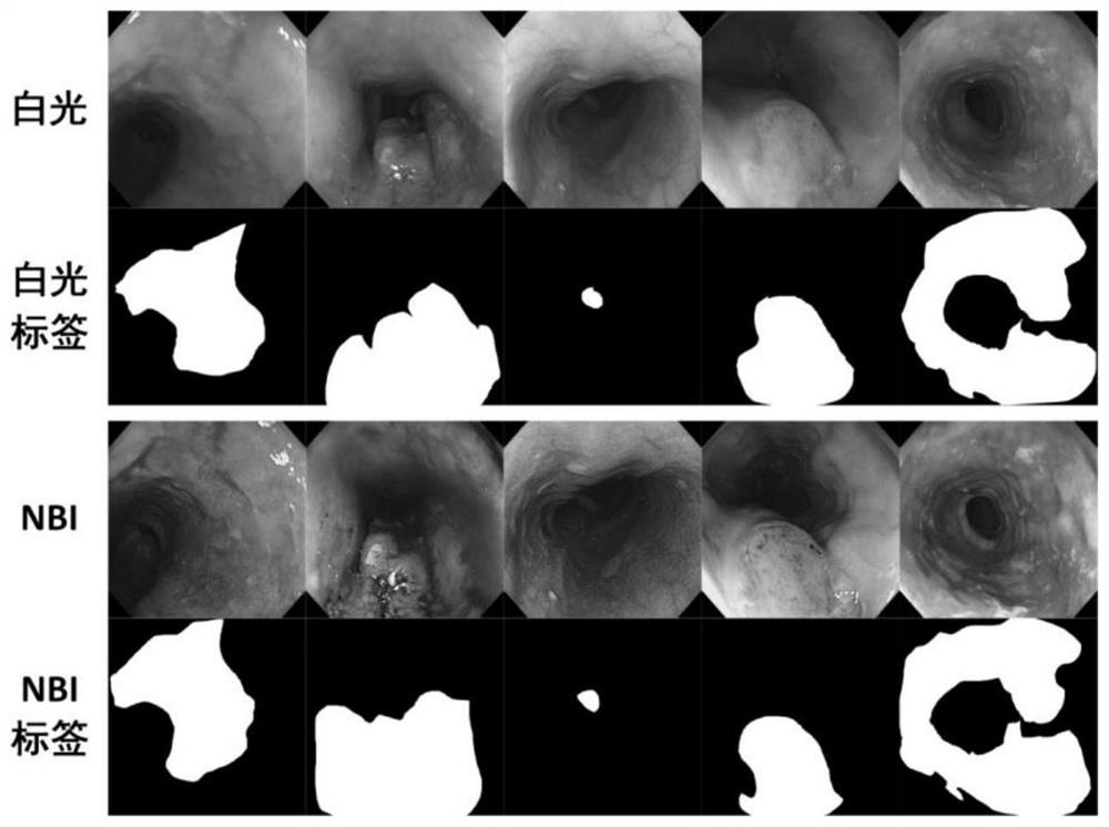
[0039] Figure 4 Showing the segmentation results...
PUM
 Abstract
Description
Claims
Application Information
 - R&D
- Intellectual Property
- Life Sciences
- Materials
- Tech Scout
- Unparalleled Data Quality
- Higher Quality Content
- 60% Fewer Hallucinations
Browse by: Latest US Patents, China's latest patents, Technical Efficacy Thesaurus, Application Domain, Technology Topic, Popular Technical Reports.
© 2025 PatSnap. All rights reserved.Legal|Privacy policy|Modern Slavery Act Transparency Statement|Sitemap|About US| Contact US: help@patsnap.com


