Ultrasonic scanning teaching method for sacrococcygeal region spine

A sacrococcygeal and ultrasound technology, applied in the field of sacrococcygeal spine ultrasound scanning and teaching, can solve problems such as reducing the safety of clinical diagnosis and treatment, inaccurate positioning of target sections, and limited application.

Inactive Publication Date: 2021-06-04
PEKING UNION MEDICAL COLLEGE HOSPITAL CHINESE ACAD OF MEDICAL SCI
View PDF0 Cites 0 Cited by
  • Summary
  • Abstract
  • Description
  • Claims
  • Application Information

AI Technical Summary

Problems solved by technology

This leads to disorder in the scanning process and inaccurate positioning of the target section when the clinician is learning to perform the ultrasound scan of the sacrum. It al

Method used


Image

  • Ultrasonic scanning teaching method for sacrococcygeal region spine
  • Ultrasonic scanning teaching method for sacrococcygeal region spine
  • Ultrasonic scanning teaching method for sacrococcygeal region spine

Examples


Embodiment Construction

[0020] In order to better understand the purpose, structure and function of the present invention, a teaching method for ultrasonic scanning of the sacrococcygeal spine of the present invention will be further described in detail below in conjunction with the accompanying drawings.

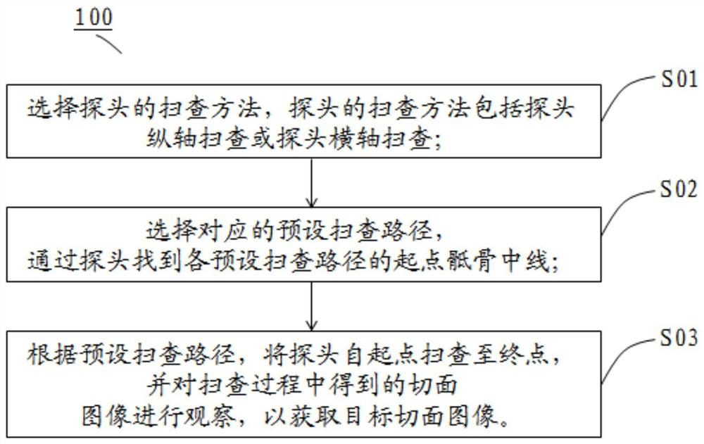
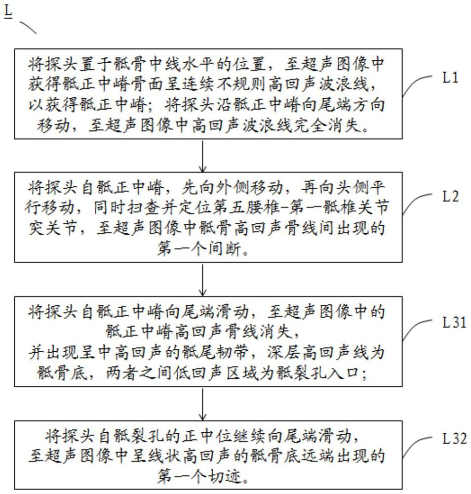
[0021] According to the teaching method 100 for ultrasonic scanning of the sacrococcygeal spine of the present invention, the steps include: step one S01, select the scanning method of the probe, the scanning method of the probe includes the longitudinal axis scanning of the probe or the horizontal axis scanning of the probe; step two S02 , select the corresponding preset scanning path L, find the sacral midline of the starting point of each preset scanning path L through the probe; step 3 S03, scan the probe from the starting point to the end point according to the preset scanning path L, and scan The section images obtained during the inspection process are observed to obtain the target section i...


PUM


Abstract

The invention belongs to the field of ultrasonic scanning teaching methods, and discloses an ultrasonic scanning teaching method for a sacrococcygeal region spine. The method comprises the following steps: step 1, selecting a probe scanning method, which comprises probe longitudinal axis scanning or probe transverse axis scanning; step 2, selecting corresponding preset scanning paths, and finding a starting point sacrum center line of each preset scanning path through a probe; and step 3, according to a preset scanning path, scanning the probe from a starting point to an end point, and observing a section image obtained in the scanning process to obtain a target section image. According to the method disclosed by the invention, the scanning path diagram of the sacrococcygeal region spine is established, so that scanning guidance for clinicians is facilitated.

Description

technical field [0001] The invention belongs to the field of ultrasonic scanning teaching methods, and in particular relates to a teaching method for ultrasonic scanning of sacral and coccygeal vertebrae. Background technique [0002] The spine of the human body includes cervical vertebrae (usually represented by "C" in medicine), thoracic vertebrae (usually represented by "T" in medicine), lumbar vertebrae (usually represented by "L" in medicine) and sacrococcygeal vertebrae (sacral tail spine, medically Usually denoted by "S"). Among them, the sacrum is composed of the sacrum (fused with 5 sacral vertebrae) and the coccyx (fused with 3-5 sacral vertebrae). The front and back sides are in an inverted triangle shape, forming the posterior upper wall of the pelvic cavity. The lower end is the tip of the sacrum, which is connected with the coccyx. The pelvic surface of the sacrum is concave and the dorsal side is kyphotic to increase pelvic volume. The probe usually include...

Claims


Application Information

Patent Timeline
IPC IPC(8): G09B23/28
CPCG09B23/286
Inventor 崔旭蕾费昱达阮侠袁青张秀华黄宇光
Owner PEKING UNION MEDICAL COLLEGE HOSPITAL CHINESE ACAD OF MEDICAL SCI
Why Eureka
Try Eureka