Neck spine ultrasonic scanning teaching method

A spine and ultrasound technology, applied in the field of cervical spine ultrasound scanning teaching, can solve the problems of disordered scanning process, long learning curve for clinicians, and inconvenience.

Inactive Publication Date: 2021-06-04
PEKING UNION MEDICAL COLLEGE HOSPITAL CHINESE ACAD OF MEDICAL SCI
View PDF0 Cites 0 Cited by
  • Summary
  • Abstract
  • Description
  • Claims
  • Application Information

AI Technical Summary

Problems solved by technology

This leads to disorder in the scanning process and inaccurate positioning of the target section for clinicians to learn to perform ultrasound scans of the cervical spine. It also leads to problems such as a long learning curve for clinicians, which makes it difficult for clinicians to learn spine ultrasound scans, and the application is therefore limited

Method used


Image

  • Neck spine ultrasonic scanning teaching method
  • Neck spine ultrasonic scanning teaching method
  • Neck spine ultrasonic scanning teaching method

Examples


Embodiment Construction

[0030] In order to better understand the purpose, structure and function of the present invention, a method for teaching cervical spine ultrasound scanning of the present invention will be further described in detail below in conjunction with the accompanying drawings.

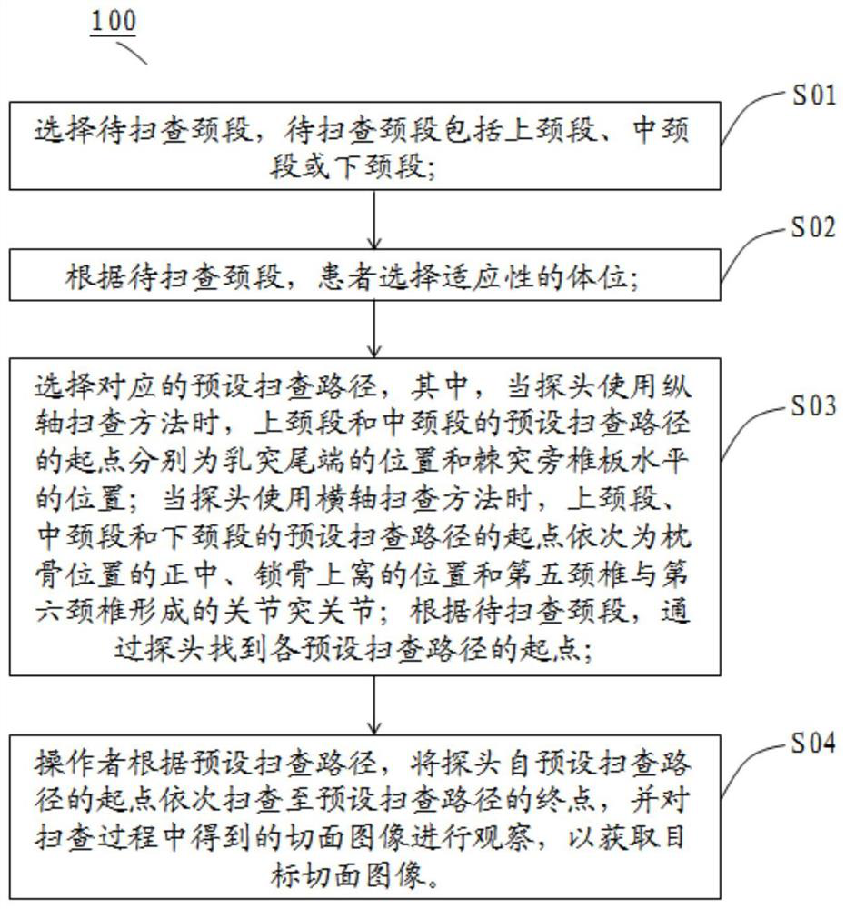
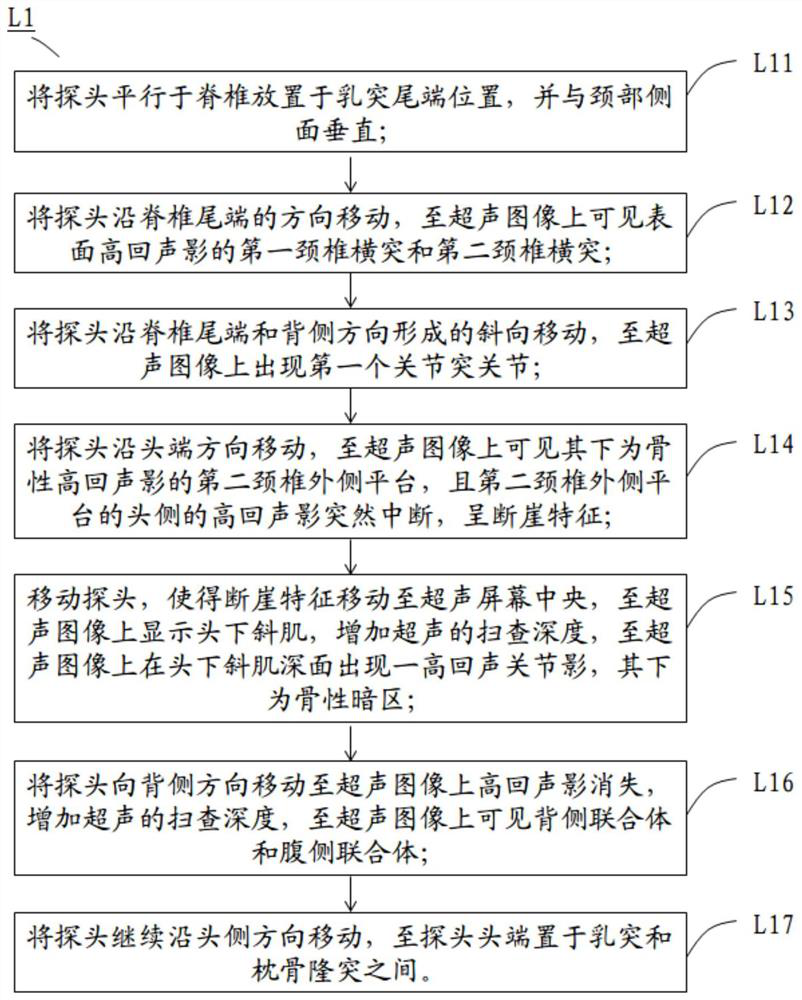
[0031] figure 1 A flow chart of a teaching method 100 for cervical spine ultrasound scanning according to the present invention is shown. Such as figure 1As shown, the steps of the cervical spine ultrasonic scanning teaching method 100 include: Step 1 S01, select the cervical segment to be scanned, and the cervical segment to be scanned includes the upper cervical segment, middle cervical segment or lower cervical segment; Step 2 S02, According to the cervical segment to be scanned, the patient selects an adaptive body position; Step 3 S03, selects the corresponding preset scanning path L, wherein, when the probe uses the longitudinal axis scanning method, the preset position of the upper cervical segment and...


PUM


Abstract

The invention belongs to the field of ultrasonic scanning teaching methods, and discloses a neck spine ultrasonic scanning teaching method. The method comprises the following steps: selecting a neck segment to be scanned; selecting an adaptive body position by a patient; selecting a corresponding preset scanning path, wherein when a probe uses a longitudinal axis scanning method, the starting points of the preset scanning paths of the upper neck section and the middle neck section are the position of the mastoid tail end and the horizontal position of a vertebral plate beside the spinous process respectively, and when the probe uses a horizontal axis scanning method, the starting points of the preset scanning paths of the upper neck section, the middle neck section and the lower neck section are the center of the occipital bone position, the position of the supraclavicular fossa and the zygopophyseal joint formed by the fifth cervical vertebra and the sixth cervical vertebra in sequence; and finding a starting point of each preset scanning path through the probe, scanning the probe from the starting point to an end point, and observing a section image obtained in the scanning process to obtain a target section image. According to the method, the scanning path diagram is established, so that ordered and perfect scanning guidance for clinicians is facilitated.

Description

technical field [0001] The invention belongs to the field of ultrasonic scanning teaching methods, in particular to a cervical spine ultrasonic scanning teaching method. Background technique [0002] The spine of the human body includes cervical spine (neck spine, usually represented by "C" in medicine), thoracic spine (usually represented by "T" in medicine), lumbar spine (usually represented by "L" in medicine) and sacral vertebrae (commonly represented by "L" in medicine) Usually denoted by "S"). Among them, the cervical spine includes seven cervical vertebrae connected in sequence, including from top to bottom: the first cervical vertebra (atlas C1), the second cervical vertebra (axis C2), the third cervical vertebra (C3) to the sixth cervical vertebra (C6) And the seventh cervical vertebra (vertebrae C7). The first cervical vertebra (atlas) and the second cervical vertebra (axis) constitute the upper cervical segment, the third to sixth cervical vertebra constitute th...

Claims


Application Information

Patent Timeline
Patent Type & Authority Applications(China)
IPC IPC(8): G09B23/28
CPCG09B23/286
Inventor 崔旭蕾费昱达申乐陈思唐帅黄宇光
Owner PEKING UNION MEDICAL COLLEGE HOSPITAL CHINESE ACAD OF MEDICAL SCI
Why Eureka
Try Eureka